 |
|
| Código de la Imagen : 3677 | |
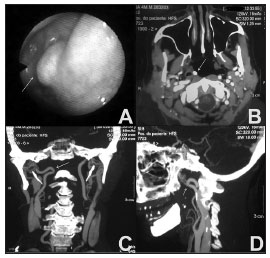
| Figure 1. A: Nasal endoscopy image showing a pulsating mass in the lateral recess of the nasopharynx. | |
Imagen publicada en: |
|
|
|
| 1 | |
| Descripción: | |
|
|
| Autor (es) del artículo de origen: | |
| Amaury de Machado Gomes; Otavio Marambaia dos Santos; Pablo Pinillos Marambaia; Carlos Augusto de Carvalho Carrera; Leonardo Marques Gomes | |
| Título y link del artículo: | |
| Anatomic variant of the internal carotid artery in the pharynx | |
| oldfiles.bjorl.org/conteudo/acervo/acervo_english.asp?id=4535 |
All rights reserved - 1933 /
2025
© - Associação Brasileira de Otorrinolaringologia e Cirurgia Cérvico Facial

