|

| Código de la Imagen : 3668 | |
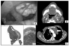
| Figure 1. A: macroscopic view of a tumor in the left tonsillar bed | |
Imagen publicada en: |
|
|
|
| 1 | |
| Descripción: | |
|
|
| Autor (es) del artículo de origen: | |
| Helena Hotz Arroyo1; Jefferson Takehara2; Allex Itar Ogawa3; Ronaldo Frizzarini5; Rui Imamura4; Henrique Moura de Paula5 | |
| Título y link del artículo: | |
| Small cell lung carcinoma metastasis to palatine tonsils | |
| oldfiles.bjorl.org/conteudo/acervo/acervo_english.asp?id=4508 |
All rights reserved - 1933 /
2026
© - Associação Brasileira de Otorrinolaringologia e Cirurgia Cérvico Facial