 |
|
| Código de la Imagen : 3667 | |
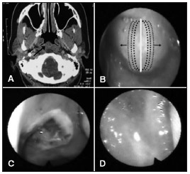
| Figure 1. A: CT scan of the sinuses showing a lesion in the nasopharynx | |
Imagen publicada en: |
|
|
|
| 1 | |
| Descripción: | |
|
|
| Autor (es) del artículo de origen: | |
| Marco Antonio Thomas Caliman1; Erika Mucciolo Cabernite2; Juliana Tichauer Vieira3; Diogo Carvalho Pasin3; Denilson Storck Fomin4 | |
| Título y link del artículo: | |
| Thornwaldt cyst - treatment with diode laser | |
| oldfiles.bjorl.org/conteudo/acervo/acervo_english.asp?id=4507 |
All rights reserved - 1933 /
2025
© - Associação Brasileira de Otorrinolaringologia e Cirurgia Cérvico Facial

