|

| Código de la Imagen : 3606 | |
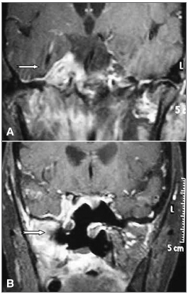
| Figure 1. A: Preoperative skull MRI (contrast-enhanced T1). | |
Imagen publicada en: |
|
|
|
| 1 | |
| Descripción: | |
|
|
| Autor (es) del artículo de origen: | |
| Bernard Soccol Beraldin1; Alexandre Felippu2; Fabio Martinelli3; Henrique Candeu Patricio3 | |
| Título y link del artículo: | |
| Tolosa-Hunt syndrome mimicking cavernous sinus tumor | |
| oldfiles.bjorl.org/conteudo/acervo/acervo_english.asp?id=4434 |
All rights reserved - 1933 /
2026
© - Associação Brasileira de Otorrinolaringologia e Cirurgia Cérvico Facial