|

| Código de la Imagen : 3532 | |
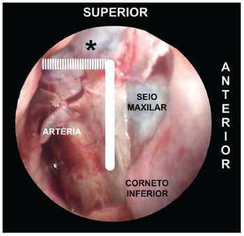
| Picture of a left nasal fossa | |
Imagen publicada en: |
|
|
|
| 4 | |
| Descripción: | |
|
|
| Autor (es) del artículo de origen: | |
| Gustavo Lara Rezende1; Vitor Yamashiro Rocha Soares2; Waldete Cabral Moraes3; Carlos Augusto Costa Pires de Oliveira4; Márcio Nakanishi5 | |
| Título y link del artículo: | |
| The sphenopalatine artery: a surgical challenge in epistaxis | |
| oldfiles.bjorl.org/conteudo/acervo/acervo_english.asp?id=4322 |
All rights reserved - 1933 /
2026
© - Associação Brasileira de Otorrinolaringologia e Cirurgia Cérvico Facial