INTRODUCTIONSince the 1980s, computed tomography (CT) has been recommended as the gold standard for assessing rhinosinusitis (RS);1-3 it is an important component in the routine approach to the chronic form of this disease.4 The advantages of CT are its high sensitivity for inflammation (opacification)5 of the paranasal sinuses (PNS) and ability to demonstrate in detail the bony labyrinth of this area, including the narrow drainage pathways of the ostiomeatal complex (OMC).6 Thus, at the same time it discards other conditions that simulate chronic rhinosinusitis (CRS), CT may reveal eventual structural obstructive factors4 that help maintain the clinical picture.7
With the advent of functional endoscopic surgery (FES), CT has become as a "map" for surgeons by demonstrating the anatomy and its variants, and the distribution of opacification.4,7,8 In effect, there has been a search for measurement tools to translate the amount of rhinosinusal opacification into numbers or degrees at CT - the so-called scores.9-15 In objectively quantifying sinus opacification, scores have made it easier to correlate CT with clinical and endoscopic parameters, which may potentially help select patients that would benefit most from FES.8,16,17
In 1997, following a comparative analysis of eight better known scores, the American Academy of Otorhinolaryngology and Head & Neck Surgery, recommended the Lund-Mackay system (LMS).18 The LMS consists of checking 0 to 2 points for each cavity (maxillary, frontal, sphenoid, anterior ethmoid and posterior ethmoid) and for the OMC, that is, six sites in each side. Zero means a normal sinus, 1(one) partial opacification, and 2(two) complete opacification. The results is the sum of scores for each side, which ranges from zero to 24.12
The LMS has been proved to be practical and accurate in subsequent studies.16,18-20 However, it has not correlated regularly with the intensity of preoperative symptoms or the degree of clinical improvement after FES.8,17,21-23 One of its caveats is that the LMS groups together any partial opacification under a single score (one), thereby attributing equal values to both the presence of fluid and mucous thickening, which have different clinical outcomes.24,25 Furthermore, studies of scores have been made mostly in adults,8,16,17,19-21 and appear not to have taken into account the effect of absent sphenoid and frontal sinuses - a common feature of patients under 12 years of age - on their results. Given that the LMS is based on the sum of scores for each rhinosinusal compartment, absent (undeveloped) sphenoid and frontal sinuses reduce the score amplitude to 16 from 24 points, which results in artificially underestimating disease and therefore a bias when applied to children.
It is hoped that a tomographic score expressing an estimate (or percentage) of the proportion of opacification within cavities may be less prone to developmental interferences, and therefore applicable indistinctly to any age group. Such as score, named the opacification/development ratio (ODR), was used by the authors in a previous study of asymptomatic children and adolescents.26 This study aimed to verify the precision and accuracy of ODR and to compare it with the LMS, for validation.
SUBJECTS AND METHODSStudy DesignA contemporary cross-sectional cohort study to assess the precision and accuracy of a diagnostic tool.
SubjectsAn evaluation was made of the exams of patients aged from 2 to 18 years with a clinical diagnosis of RS that had been referred to the radiology unit for CT of the PNS from April 2002 to July 2004. Clinical diagnostic criteria, classification and intensity of disease were not arbitrated, as these exams served only as samples for repeated measures of sinus opacification on CT. Subjects with technically imperfect exams that did not permit an adequate appraisal of all PNS were excluded. Only the first exam was included in patients with two exams.
METHODSExams were carried out using a Toshiba X-vision (Toshiba, Tokyo, Japan) device; sequential 1 to 2mm coronal sections, eventually associated with axial sections, were done; no endovenous contrast media were used. Children under the age of 4 years were generally anesthetized for the exam; these patients were placed in dorsal decubitus, with the neck hyper-extended, and the gantry angulated to keep the coronal plane as the reference. No exam had its technical or operating conditions altered because of this study. The images were recorded in 1500-2500 opening and 100-400 level windows.
The ODR (Frame 1) separately assesses development and opacification. All sinuses and OMCs are considered as pairs, one for each side. The Frame below shows development scores for each sinus as 3(three) if present and 0(zero) if absent. The sum of five sites (4 cavities and the OMC) may reach 15 points for each side (30 in total) if all sinuses are developed. For opacification (see column in the Frame below), each cavity scores from 0 to 3 according to the opacified area: normal = 0(zero); < 2/3 = 1(one);
> 2/3 = 2(two); total = 3(three). Here, the OMC scores zero if normal, or 3 if opacified. Complete opacification of all sites in both sides adds up to 30 points. ODR calculation consists of the ratio: "sum of right and left opacification" (numerator) / "sum of right and left development" (denominator). The results ranges from 0 (normal) to 1 (complete opacification of the developed area). When multiplied by 100, it yields an estimated percentage of the global opacified area. The LMS was applied as previously described.12,18

Frame 1. Formula for the opacification/development ratio (ODR).
Based on the two methods (ODR and LMS), two radiologists analyzed the images twice, totaling four sessions, separated by at least two weeks. Examiners had no access to each other's results or to their own first assessments.
After verifying the mean values of four LMS readings (two from each examiner), the sample was classified into opacification categories (normal, mild, moderate or severe), where 0 (zero) was normal, 0 to 3 was mild, 4 was 10 was moderate, and over 10 was severe. This division was based on studies by Bhattacharyya and Fried16 who, based on an accuracy analysis, defined an LMS
> 4 as an appropriate cut-off point for defining a positive CT for RS. In this same paper, the group of patients with clinically diagnosed CRS had a mean LMS score ≈ 10 (Frame 2). A preliminary analysis of the sample distribution revealed that each of these four categories (normal, mild, moderate or severe) contained about 25% of the sample (quartiles). Thus, quartile points on the scale were set to extract equivalent ODR borderline values; values > 15 were moderate, and values > 50 were severe (Frame 2).

Frame 2. Classification parameters of the sample by categories for each score
This study design was approved by a CONEP-registered institutional review board, and registered under the protocol number 0814.0.146.000-08. A free informed consent form was made available beforehand to patients or their caretakers to authorize their participation in the study.
The Statistical Package for the Social Sciences (SPSS version 13.0; SPSS Inc., Chicago, IL, USA) was used for the statistical analysis. Method accuracy (reproducibility) was assessed using the intra and interobserver agreement Kappa coefficient (k) for categorical variables. Intra and interclass coefficients (intra-CC and inter-CC) were applied to assess agreement among quantitative variables (numerical scales of scores). Pearson's coefficient was applied to verify linear correlation between scales of both methods. ODR accuracy was extracted taking the LMS as the gold standard, with receiver operating characteristic (ROC) curve analysis.
RESULTSThere were 81 exams from different patients; 17 were excluded because of incomplete images or artifacts. Of the 64 remaining exams, 38 were from males (60.3%). Ages ranged from 2 to 18 years (mean - 10 years, SD=4). Two patients had undergone prior rhinosinusal surgery.
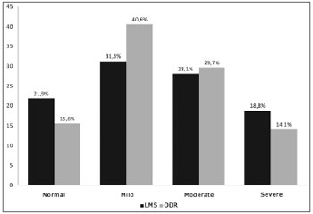
The prevalence of exams with abnormalities was 78.1% (LMS) and 74.4 (ODR). Scoring of four sets of readings ranged from 0 to 88, mean 21.3 (SD=23.1), for the ODR. The variation was 0 to 20, mean 5.2 (SD=5.3), for the LMS. Chart 1 shows the sample distribution by category, according to each score.

Chart 1. Distribution of the sample per category for each score (%). - LMS: Lund-Mackay system; ODR: opacification/development ratio
Table 1 shows the intra and interobserver agreement indices based on the ODR and LMS Kappa (k) coefficients, based on a classification by category. The ODR intraobserver was calculated using a single value per exam, which was extracted from the arithmetic mean of two readings for each examiner. The same was done for the LMS. Tables 2 and 3 show the variability calculations of score measurements based on the intra and interclass coefficients (inter-CC and intra-CC),27 which correspond respectively to the intraobserver and interobserver variability. In this test, the reproducibility coefficient (r) is an estimate of the maximum difference that could be obtained between two random measures of the same subjects; it is the limit within which are 95% of the differences. The intra-CC r was not more than 4.2 (0 to 24 scale) for the LMS; it was not more than 15.4 (0 to 100 scale) for the ODR. The inter-CC r was 4.0 (LMS) and 15.6 (ODR).
Each exam was represented by a single ODR value and a single LMS value extracted from the mean of four readings (two by each examiner) for the correlation between methods. The methods agreed substantially for categorizing the disease (normal, mild, moderate and severe), where k = 0.68 for the examiner L.F.F., and k=0.76 for the examiner S.A.A.N (p < 0.001). Chart 2 shows the linear correlation between ODR (0 to 100) and LMS (0 to 24) quantitative scales. Pearson's coefficient revealed a strong linear correlation (r=0.97) between methods. The ODR score may be converted to its LMS equivalent with the formula: LMS = 0.22 x ODR + 0.43 (r2=0.95).

Chart 2. Linear regression between scales of two scores LMS: Lund-Mackay system; ODR: opacification/development ratio. Pearson's coefficient (r=0.97). LMS=0.22 x ODR + 0.43 (r2=0.95).
ODR accuracy was calculated taking the LMS as a reference test (positive when the LMS
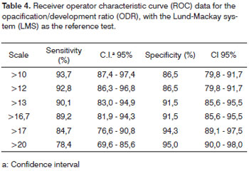
> 4). Inclination of the ROC curve (Chart 3) indicates good accuracy. Table 4 shows the numbers extracted from the curve, where the best ODR sensitivity and specificity values were between 13 and 16.7.

Chart 3. Receiver Operator Characteristic curve for the opacification/development ratio. Lund-Mackay system (LMS) as the reference test (positive = 4). - ODR: opacification/development ratio

In the present study, the prevalence of exams with any opacification (74% to 78%) was on average similar to the values reported by other authors in disease populations.11,16,25,28,29
The mean LMS score was 5. Reported numbers in the literature vary depending on the sample population. Studies of patients undergoing surgery for the treatment of CRS - patients for which medical treatment was insufficient - resulted in higher means (9 to 13)16,22 and were more intensely altered30 compared to those shown in Chart 1. On the other hand, results are closer in studies of subjects undergoing primary evaluation of RS. A study with this type of population gathered data from several North-American centers23 and found that the LMS mean ranged from 1(one) to 5(five). This population was probably more similar to our series, as our inclusion criteria for exams were intentionally deliberately ample, regardless of the intensity or duration of the clinical picture. Thus, many patients were not necessarily FES candidates, and even subjects with mild or self-limited symptoms were likely to be included.
The ODR mean was 21. There are no parameters from diseased populations in the literature for comparison purposes. A single previous study of ODR applied this method to evaluated asymptomatic children undergoing studies of the cranium unrelated to RS to seek incidental sinusal findings.26 In this study, the ODR mean was lower (15), as expected for individuals aged 3 years and above, which was similar to our age range.
General ODR inter and intraobserver agreement indices for classifying patients into categories were comparable, and at times superior, to the LMS; k generally remained between 0.6 and 0.9, which is considered as substantial agreement.31 Oluwole et al.20 found a similar LMS performance (interobserver - 0.72; intraobserver - 0.73), which are on average superior to those found using other methods such as in Jorgensen,10 May13 and Newman;14 their intra and interobserver k variables ranged from 0.34 to 0.66. Analysis of quantitative score scales showed that ODR intra and interclass coefficients had significant intraobserver and interobserver reproducibility values (> 0.90), which were invariably higher than LMS values.
There was a strong linear correlation between methods, suggesting that the ODR responds to sinus opacification intensity similarly to the LMS. There was also substantial agreement in sample categorization when both methods were compared, meaning that the cut-of points classified similarly the intensity of opacification.
Nearly all LMS and ODR correlation showed a trend towards higher agreement coefficients in the "normal" and "severe" categories compared to the mild and moderate categories, suggesting that the limit between intermediate categories may be inherently difficult to establish using CT when writing its report, or that the cut-off points for these categories require fine tuning.
The ODR accuracy was calculated indirectly, taking the LMS as the gold standard (positive
> 4). The ROC curve was accurate, with best results in the 13 to 17 ODR range. It should be noted that lower values do not necessarily discard inflammation. Likewise, a positive test should not be used alone to establish a diagnosis of RS. Due correlation with clinical findings should be the most important guiding factor.4,32
The LMS was used here for comparison because it is currently the most widely accepted score in the academic community. Some of its deficiencies, however, have been discussed openly in its recommending text.18 It has been suggested that partial opacification could be partitioned into more points, rather than just one, to better differentiate intermediate cases. This same article comments that hypoplastic frontal sinuses in adults should score zero in the LMS. There are not references by the LMS authors about what to do in cases of undeveloped sinuses in children. Scoring zero for undeveloped sinuses, was done in a study of children in Oceania,33 certainly does not solve underestimation of disease.
Although FES has fewer indication in children and adolescents compared to adults, and is used as the last measure in chronic refractory cases, this procedure has yielded satisfactory results in this age group,34 which underlines the importance of an appropriate tomographic score.
None of the currently used scoring systems provide a final result that conveys an idea of opacified area proportion, as the ODR. A few generate categorical variables (groups),9,11,13,15 while others - Jorgensen,10 Newman et al.,14 and the Miami University method30 - are similar to the LMS; they apply points according to the degree of opacification in each sinus. Because of their features, incomplete development may affect all of these systems.
Some authors have studied incidental tomographic findings in the PNS of children without sinus disease and written their own quantification criteria of sinus opacification; these contain the idea of proportion for opacification. Manning et al.35 attributed opacification intensity categories in degrees, but the final result was given as classes, which did not translate the total area of disease involvement. Lesserson et al.36 and Diament et al.37 applied similar criteria, but made a similar decision when presenting the final score (classes).
Because it is a representation of opacification percentages, we expect that ODR results are less affected by the number or size of developed cavities, compared to the LMS. Subsequent studies should include a sufficiently large sample aged below 12 years to confirm this hypothesis. Our sample of subjects with incompletely developed sinuses was small and did not allow consistent statistics.
Our objectives did not require prospective control of diagnostic criteria for RS, as it did not involve correlating tomographic and clinical findings. The exam sample served only for repeated measures of tomographic abnormalities to provide data so that we could assess inter and intraobserver variations of the ODR. In this context, it is desirable to include a wide range of clinical states, from oligosymptomatic to rich clinical pictures, so that the full amplitude of scores is represented, from zero (normal exam) to maximum degrees of opacification. ODR accuracy (sensitivity and specificity) was measured based on LMS as a reference for the same sample. Thus, it is unlikely that lack of data would have affected our results.
Nevertheless, signs and symptoms are currently used as the best parameter for diagnosing RS and measuring the response to therapy. Thus, it is essential for subsequent studies to directly evaluate the accuracy of ODR relative to clinical findings; this will require a diseased population and controls (possibly with CT of the orbit, which is technically similar to examination of the PNS). In this study design, signs and symptoms should be controlled prospectively and with rigor, preferably using a clinical scoring systems. Other parameters may be used as references, such as endoscopic findings or analysis of sinus puncture material.
Patients operated previously were not excluded, as anatomical peculiarities were not expected to affect ODR inter or intraobserver variability; it would be desirable to separate these patients in a sample of prevalence studies or when clinical or surgical data are used as parameters.
CONCLUSIONThe opacification/development ratio (ODR) is an accurate method for evaluating rhinosinusitis in children and adolescents; it is precise and correlates strongly with the Lund-Mackay system (LMS).
The ODR may predict the LMS value with the formula LMS=0.22 x ODR + 0.43.
The sensitivity and specificity of an ODR value of 15 as a cut-of point for a positive test was close to 90%, based on the LMS as the gold standard.
REFERENCES1. Zinreich SJ, Kennedy DW, Rosenbaum AE, Gayler BW, Kumar AJ, Stammberger H. Paranasal sinuses: CT imaging requirements for endoscopic surgery. Radiology. 1987;163(3):769-75.
2. Diament MJ. The diagnosis of sinusitis in infants and children: x-ray, computed tomography, and magnetic resonance imaging. Diagnostic imaging of pediatric sinusitis. J Allergy Clin Immunol. 1992;90(3 Pt 2):442-4.
3. McAlister WH, Parker BR, Kushner DC, Babcock DS, Cohen HL, Gelfand MJ et al. Sinusitis in the pediatric population. American College of Radiology. ACR Appropriateness Criteria. Radiology. 2000;215 Suppl:811-8.
4. Rosenfeld RM, Andes D, Bhattacharyya N, Cheung D, Eisenberg S, Ganiats TG et al. Clinical practice guideline: adult sinusitis. Otolaryngol Head Neck Surg. 2007;137(3 Suppl):S1-31.
5. Zinreich SJ. Rhinosinusitis: radiologic diagnosis. Otolaryngol Head Neck Surg. 1997;117(3 Pt 2):S27-S34.
6. Laine FJ, Smoker WR. The ostiomeatal unit and endoscopic surgery: anatomy, variations, and imaging findings in inflammatory diseases. AJR Am J Roentgenol. 1992;159(4):849-57.
7. Kennedy DW, Zinreich SJ, Rosenbaum AE, Johns ME. Functional endoscopic sinus surgery. Theory and diagnostic evaluation. Arch Otolaryngol. 1985;111(9):576-82.
8. Hopkins C, Browne JP, Slack R, Lund V, Brown P. The Lund-Mackay staging system for chronic rhinosinusitis: how is it used and what does it predict? Otolaryngol Head Neck Surg. 2007;137(4):555-61.
9. Friedman WH, Katsantonis GP, Sivore M, Kay S. Computed tomography staging of the paranasal sinuses in chronic hyperplastic rhinosinusitis. Laryngoscope. 1990;100(11):1161-5.
10. Jorgensen RA. Endoscopic and computed tomographic findings in ostiomeatal sinus disease. Arch Otolaryngol Head Neck Surg. 1991;117(3):279-87.
11. Kennedy DW. Prognostic factors, outcomes and staging in ethmoid sinus surgery. Laryngoscope. 1992;102(12 Pt 2 Suppl 57):1-18.
12. Lund VJ, Mackay IS. Staging in rhinosinusitus. Rhinology. 1993;31(4):183-4.
13. May M, Levine HL, Schaitkin B. Results of surgery. In: Levine HL, May M, editors. Rhinology and sinusology. New York: Thieme Medical Publishers, Inc., 1993:176-192.
14. Newman LJ, Platts-Mills TA, Phillips CD, Hazen KC, Gross CW. Chronic sinusitis. Relationship of computed tomographic findings to allergy, asthma, and eosinophilia. JAMA. 1994;271(5):363-7.
15. Metson R, Gliklich RE. Computed tomography to evaluate chronic sinusitis. JAMA. 1994;272(11):852.
16. Bhattacharyya N, Fried MP. The accuracy of computed tomography in the diagnosis of chronic rhinosinusitis. Laryngoscope. 2003;113(1):125-9.
17. Bhattacharyya N. Radiographic stage fails to predict symptom outcomes after endoscopic sinus surgery for chronic rhinosinusitis. Laryngoscope. 2006;116(1):18-22.
18. Lund VJ, Kennedy DW. Staging for rinosinusitis. Otolaryngol Head Neck Surg.1997;117(Suppl):S35-S40.
19. Metson R, Gliklich RE, Stankiewicz JA, Kennedy DW, Duncavage JA, Hoffman SR et al. Comparison of sinus computed tomography satging systems. Otolaryngol Head Neck Surg. 1997;117:372-9.
20. Oluwole M, Russell N, Tan L, Gardiner Q, White P. A comparison of computerized tomographic staging systems in chronic sinusitis. Clin Otolaryngol Allied Sci. 1996;21(1):91-5.
21. Sharp HR, Rowe-Jones JM, Mackay IS. The outcome of endoscopic sinus surgery: correlation with computerized tomography score and systemic disease. Clin Otolaryngol Allied Sci. 1999; 4(1):39-42.
22. Bradley DT, Kountakis SE. Correlation between computed tomography scores and symptomatic improvement after endoscopic sinus surgery. Laryngoscope. 2005;115(3):466-9.
23. Stewart MG, Sicard MW, Piccirillo JF, Diaz-Marchan PJ. Severity staging in chronic sinusitis: are CT scan findings related to patient symptoms? Am J Rhinol. 1999;13(3):161-7.
24. Wald ER. Radiographic sinusitis: illusion or delusion? Pediatr Infect Dis J. 1993;12(9):792-3.
25. Dutra LD, Marquiori E. Tomografia Computadorizada helicoidal dos seios paranasais na criança: avaliação das sinusopatias inflamatórias. Radiol Bras. 2002; 35(3):161-9.
26. Araújo Neto S, Souza A, Pereira I, Baracat E. Alterações incidentais dos seios da face na tomografia computadorizada do crânio e órbitas em crianças. Radiol Bras. 2005;38(4):245-50.
27. Bland JM, Altman DG. Statistical methods for assessing agreement between two methods of clinical measurement. Lancet. 1986;1(8476):307-10.
28. Garcia DP, Corbett ML, Eberly SM, Joyce MR, Le HT, Karibo JM et al. Radiographic imaging studies in pediatric chronic sinusitis. J Allergy Clin Immunol. 1994;94(3 Pt 1):523-30.
29. Lindbaek M, Johnsen UL, Kaastad E, Dolvik S, Moll P, Laerum E et al. CT findings in general practice patients with suspected acute sinusitis. Acta Radiol. 1996;37(5):708-13.
30. Lehman DA, Casiano RR, Polak M. Reliability of the University of Miami chronic rhinosinusitis staging system. Am J Rhinol. 2006;20(1):11-9.
31. Landis JR, Koch GG. The measurement of observer agreement for categorical data. Biometrics. 1977;33:159-74.
32. Subcommittee on Management of Sinusitis and Committee on Quality Improvement. Clinical practice guideline: management of sinusitis. Pediatrics. 2001;108(3):798-808.
33. Lim WK, Ram B, Fasulakis S, Kane KJ. Incidental magnetic resonance image sinus abnormalities in asymptomatic Australian children. J Laryngol Otol. 2003;117(12):969-72.
34. Bernal-Sprekelsen M, Massegur SH, Tomas BM. Paediatric endoscopic sinus surgery (PESS): review of the indications. Rev Laryngol Otol Rhinol (Bord ). 2003;124(3):145-50.
35. Manning SC, Biavati MJ, Phillips DL. Correlation of clinical sinusitis signs and symptoms to imaging findings in pediatric patients. Int J Pediatr Otorhinolaryngol. 1996;37(1):65-74.
36. Lesserson JA, Kieserman SP, Finn DG. The radiographic incidence of chronic sinus disease in the pediatric population. Laryngoscope. 1994;104(2):159-66.
37. Diament MJ, Senac MO, Jr., Gilsanz V, Baker S, Gillespie T, Larsson S. Prevalence of incidental paranasal sinuses opacification in pediatric patients: a CT study. J Comput Assist Tomogr. 1987;11(3):426-31.
1. Master's degree, coordinator of the Image Diagnosis Module of the Paraiba Medical School (Faculdade de Ciências Médicas da Paraíba).
2. Doctoral degree, assistant professor, Pediatrics Department, UNICAMP Medical School.
3. Specialist, medical radiologist.
Campinas State University (Universidade Estadual de Campinas - UNICAMP)
Send correspondence to:
Severino Aires de Araujjo Neto
Rua Maria Helena Rocha 113 apto. 1102 Aeroclube
João Pessoa PB 58036-670
Paper submitted to the BJORL-SGP (Publishing Management System - Brazilian Journal of Otorhinolaryngology) on September 11, 2009; and accepted on November 27, 2009. cod. 6635


