INTRODUCTIONWaldeyer's lymphatic ring is part of the first line of defense against pathogens; it is located at the entrance point of the airways and digestive tract. It consists of lymphoid tissue, which includes the palatine and pharyngeal tonsils. Many microorganisms may infect these tissues and cause tonsillitis; the most frequent are bacteria.1 The mechanism by which some children develop recurring tonsillitis is still unclear, as is the cause of exaggerated tonsillary enlargement. Many hypotheses have been raised, such as possible resistance of microorganisms to antibiotic therapy, and Streptococcus
a interfering as an oropharyngeal protector against Streptococcus ▀ to prevent recurring tonsillitis. Some studies have suggested that virus infections may be involved in recurring infections, including the Epstein-Barr virus (EBV) and herpes simplex viruses.2-4
The EBV was discovered in 1964 in a culture of Burkitt's lymphoma cells; in 1968 studies revealed that it caused infectious mononucleosis.5 It is a herpesviridae of the subfamily gamaherpesvirinae, which infects most individuals before adult life. During the first infection, viruses are transmitted by saliva and invade the oropharyngeal epithelial cells, which are destroyed; it then infects circulating B lymphocytes, within which the virus becomes latent.4 The EBV genome consists of a linear 172 kilobase DNA molecule coding about 100 viral proteins; however, only 10 genes are expressed in vitro in infected B lymphocytes (latency). These 10 genes consist of six nuclear proteins (EBNAs 1, 2, 3A, 3B, 3C and EBNA-LP), two membrane proteins (LMP-1 and LMP-2), and two small RNAs (EBER 1 and EBER 2).6 The nuclear antigen protein 1 (EBNA-1) binds to the viral DNA so that the viral genome remains in infected cells as a circular episome; LMP-1 expression in immunocompromised individuals may induce B lymphocyte transformation and the onset of lymphoproliferative conditions.6
In contrast with in vitro studies, the EBV replication site has not been established in vivo. Infectious mononucleosis and oral hairy leukoplakia have been used as models for studying the replication mechanism of the virus.3 The EBV replication mode in healthy subjects is still unknown. The tonsils appear to be candidate sites for EBV replication.7,8 Babcock (1998) detected linear episomal forms of EBV DNA in tonsillary lymphocytes. These linear forms suggest that virus DNA is replicating in tonsillary lymphocytes in healthy subjects previously infected by the EBV. Until recently it was thought that the virus was capable of infecting only B lymphocytes and epithelial cells; there have, however, been reports of infected normal T lymphocytes.3
The EBV is associated not only with infectious mononucleosis but also with other benign diseases, such as oral hairy leukoplakia, and malignancies, such as Hodgkin's lymphoma, non-Hodgkin's B and T cell lymphomas, and nasopharyngeal, gastric and breast carcinomas. It is currently being associated with autoimmune diseases, such as lupus erythematosus and multiple sclerosis.5,9
Many molecular techniques are being used to demonstrate the presence of the EBV, such as the polymerase chain reaction (PCR) and in situ hybridization (IHS). PCR makes it possible to detect minimal amount of viral DNA in tissues and smears. According to Peiper (1990), PCR amplification of specific EBV genome sequences is a rapid, sensitive and specific method for identifying viral DNA. Furthermore, this method makes it possible to study biopsies that were paraffined and kept in files, which permits retrospective studies.
The purpose of this study was to investigate the association between the EBV and recurring tonsillitis by identifying the viral genome using PCR and the LMP-1 proteins by using immunohistochemistry.
MATERIAL AND METHODA cross-sectional study was undertaken for analyzing the prevalence of EBV infection in tonsillectomy specimens, in which the indication for surgery had been recurrence and significant volume increase. The Research Ethics Committee of the Faculdade de Medicina (Medical School) - CEP-CMM approved this study (number 077/06). The slides of tonsillectomies with a diagnosis of hypertrophic chronic tonsillitis were selected among the tonsillectomies studied at the Pathology Unit from 1999 to 2006. After excluding inadequate cases (insufficient material, technical artifacts, inadequate fixation) 24 paraffined blocks with tonsils fixated in 10% buffered formaldehyde remained for study.
The nested PCR reaction using DNA extracted from three 5Ám thickness tissue slices was used for identifying viral DNA. After sectioning each sample, the microtome was cleaned and the histological scalpels were changed. For DNA extraction the samples were deparaffinated in three xylene baths at 65║C and hydrated with successive baths of 100%, 95% and 70% alcohol. After centrifugation, tissues were resuspended in 220ÁL of autoclaved milli-Q water and digested at 56║C in the presence of 10% SDS (30Ál) and K proteinase at 25mg% (6ÁL). K proteinase was added each day until the samples were completely digested. The suspension was extracted with phenol/chloroform/isoamylic alcohol (25:24:1) to remove undigested proteins, and after DNA precipitation with sodium acetate (3M) (35ÁL) and absolute alcohol (1mL), the samples were centrifuged and resuspended with sterile milli-Q water (50ÁL).11 Extracted genomic DNA quality and estimated quantity was verified using 1.7% agarose gel electrophoresis containing ethidium bromide. The genomic DNA of all samples underwent ▀-globin gene PCR (constitutive cell gene) to check whether the samples contained amplifiable DNA. The nested PCR reaction to amplify the EBNA-1 gene was used for identifying the EBV DNA, generating a 279 bp fragment in the first reaction and a 209 bp fragment in the second reaction.12 The first reaction mix, which identifies the herpex virus, contained 2.5Ál of DNA from each extracted sample, 0.5Ál of primers at 50ÁM (5'AAG-GAG-GGT-GGT-TTG-GAA-AG 3' and 5'AGA-CAA-TGG-ACT-CCC-TTA-GC 3'), 2.5Ál of a buffer solution (10mM TrisHCL pH8.3, 50mM of KCL), 0.75Ál of MgCl at 2mM, 2Ál dNTP at 2mM, and 0.2Ál of Taq polymerase Invitrogen (5U/Ál), and enough milli-Q water to reach a 25 Ál end volume. The DNA used as a positive control was extracted from the EBV virus infected B9507 cell line; the negative control was done with sterile milli-Q water. Amplification was done using the following program: 2 minutes at 94║C, followed by 35 30-second cycles at 94║C, 30 seconds at 55║C, and 30 seconds at 72║C, followed by a 10 minute final extension at 72║C. For the second reaction, specific primers for the EBV (5' ATC-GTG-GTC-AAG-GAG-GTT-CC 3' and 5' ACT-CAA-TGG-TGT-AAG-ACG-AC 3') were used under the same conditions with the following program: 45 seconds at 94║C, followed by 35 20-second cycles at 94║C, 30 seconds at 55║C, 30 seconds at 72║C, followed by a 10 minute final extension at 72║C. Identification of gene amplification was done using an ethidium bromide stained 1.7% agarose gel, in which 7Ál of each nested PCR product was separated by electrophoresis; a 100 bp molecular weight was used as the parameter (Fermentas).
To investigate the presence of the LMP-1 latency protein, paraffined blocks were microtomized and 5Ám thickness sections were placed on pretreated slides with an adhesive (Silano«4%), after which deparaffining and washing with Tris-Buffered Saline was carried out. The slides were treated for antigen recovery during 30 minutes in a double boiler at 96║C with a citrate buffer. The primary antibody was the monoclonal anti-LMP-1 (DAKO«) diluted at 1:400 according to the manufacturer's instructions, and the kit LSAB (DAKO«) containing the biotinilated second antibody and the streptavidine and peroxidase conjugate. Revelation was done using a chromogenous solution (Kit DAB-DAKO«), and counterstaining was done with Harris' hematoxyllin. Sections of lymphoma and inflammatory fibrous hyperplasia were used as positive and negative controls.
RESULTSThe sample used in this study originated from pediatric patients aged from 2 to 12 years (mean 7.4 years, standard deviation 3.1 years), of which 15 (62.5%) were female and 9 (37.5%) were male. Tonsillectomy was indicated due to tonsillary hypertrophy and adenoid vegetations in 17 cases, due to tonsillary hypertrophy and adenoid vegetations and recurring tonsillitis in 5 cases, and due to recurring tonsillitis in 2 cases.
The genomic DNA visualized in agarose gel showed good quantity of DNA in extracted samples. Of 24 tonsils subjected to PCR, amplification of the ▀-globin gene was detected in 16 (66.7%) (Fig. 1). Of these, 10 (62.5%) amplified for the EBV genome (Table 1). EBV-DNA was detected in 13 (54.1%) of 24 tonsils (Fig. 2), of which 10 cases were from female patients (66.7% of 15 women) and 10 cases were from patients that comprised 58.8% of those aged from 6 to 12 years (Table 2). Investigation of the LMP-1 latency protein in 24 tonsils revealed immune reactivity in few lymphocytes of 9 tonsils (37.5%) (Fig. 3).
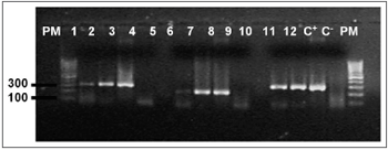
Figure 1. Agarose gel (1,7%) electrophoresis showing amplification of the
b-globin gene, (268bp) PM - Molecular weight 100bp, Samples 1 - 12, C+ - DNA extracted from the line B9507, C- - Milli-Q water.
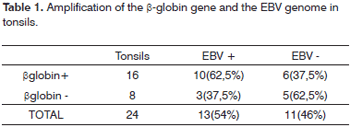
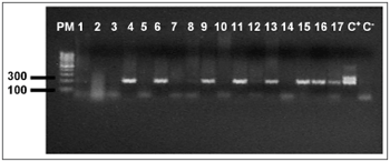
Figure 2. Agarose gel (1,7%) electrophoresis showing amplification of the EBV genome, (209bp) PM - Molecular weight 100bp, Samples 1 - 17, C+ - DNA extracted from the line B9507, C- - Milli-Q water.
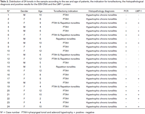
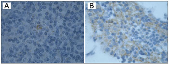
Figure 3. Immunohistochemistry showing positive results for the LMP-1 in cases A and B (1000X).
The Epstein-Barr virus causes infectious mononucleosis and oral hairy leukoplakia; it has also been associated with numerous malignancies including Hodgkin's disease, B and T lymphomas, nasopharyngeal carcinomas, and gastric carcinomas in immunosuppressed patients. Little is known, however, about the pathogeny of the virus in immunocompetent patients. Many researchers have suggested that the tonsils are a possible replication site for this virus.2,3,8,13
Molecular techniques have been used often for diagnosing and monitoring patients with virus diseases. The EBV-DNA can be identified by in situ hybridization (ISH) and the polymerase chain reaction (PCR); some authors consider both equally sensitive for detecting the EBV.14 Additionally, viral proteins found in latent and replicative infection may be identified by immunohistochemical techniques.2-4,13 The tonsils are considered the site of initial infection, and of viral persistence and replication. The EBV may infect the tonsils of children and become involved in recurring tonsillitis.2,15,16
In our study we selected the tonsils of children with a mean age of 7,4 years and a diagnosis of chronic and hypertrophic tonsillitis. We believe that the significant number of recurring tonsillary infections in childhood may be associated with the EBV; initial contact with this virus often occurs around age 7 years. Sixteen of 24 tonsils showed positive amplification for ▀-globin, thus demonstrating the presence of constitutional DNA, which was amplifiable, in the material that was extracted. In 10 of these 16, amplification of EBV was obtained, as in three tonsils that were negative for ▀-globin, suggesting that, although EBV-DNA was amplified in these three samples, we cannot state that the other five EBV-negative samples did not amplify by lack of the virus or low quantity or quality of DNA. It can be stated that the EBV infected 54.1% of the tonsils in our sample; but mapping this infection, which would make it possible to identify the target cell population, faces technical limits. Niedobitek et al.17 used the ISH to define the pattern and distribution of EBV-positive lymphoblasts found mostly in extrafollicular areas. The prevalence of EBV infection in tonsillitis varies according to the detection method. Studies using the ISH for detecting EBER found a 26%,19 a 29%,2 and a 65%4 association of the EBV with tonsillitis. Other studies using the PCR found a prevalence of up to 65% (11%,20 58%,15 and 64%17). The PCR thus confirms its higher sensitivity for molecular detection, and suggests an association between the EBV and tonsillitis.
Ping-Ching Pai et al.'s15 results using nested PCR to investigate an association between the EBV and 57 tonsillitis cases (58%) and 31 tonsils with malignancies (51%) suggest that the association is not restricted only to recurring tonsillitis.
Ikeda et al.3 (2000) used the ISH, the RT-PCR, and immunohistochemistry in 15 fragments of tonsils from patients with chronic tonsillitis. In a detailed study, these authors showed that tonsillary lymphocytes are not only a reservoir but also a replication environment for the EBV. Our results similarly found the LMP-1 (the EBV latency protein) in 37.5% of tonsils in our sample, showing that immunohistochemical methods may be an initial investigation method for latent EBV infection. Many question remain to be studies in the pathogeny of EBV infection; its association with the tonsils is particularly challenging.
CONCLUSIONIdentification of a high (54.1%) prevalence of EBV-DNA and identification of the LMP-1 (37.5%) in recurring tonsillitis in children suggest that the tonsils may be reservoir for the EBV, and that this virus may be involved in recurring infection. Many aspects of latent and replicative EBV infection remain unclear, and are possible points for future research.
REFERENCES1. Ciceran A. Manual Boca e Faringe. www.iapo.org. 2004; 84-94.
2. Endo LH, Ferreira D, Montenegro MC, Pinto GA, Altemani A, Bortoleto AE et al. Detection of Epstein-barr virus in tonsillar tissue of children and the relationship with recurrent tonsillitis. Int J Pediatr Otorhinolaryngol 2001;58:9-15.
3. Ikeda T, Kobayashi R, Horiuchi M, Nagata Y, Hasegawa M Mizuno F et al. Detection of lymphocytes productively infected with Epstein-Barr virus in non- neoplastic tonsils. J Gen Virol 2000;81:1211-6.
4. Chagas AC, Endo LH, Santos WLC, Pinto GA, Sakano E, Brousset P et al. Is there a relationship between the detection of human herpesvirus 8 and Epstein-Barr virus in Waldeyers ring tissues? Int J Pediatr Otorhinolaryngol 2006:1923-7.
5. Ribeiro-Silva AR, Zucoloto S. O papel do vÝrus Epstein-Barr na tumorigŕnese humana. Medicina RibeirŃo Preto 2003;36:16-23.
6. Cruchley AT, William DM. Epstein-barr virus: biology and disease. Oral Dis Suppl 1 1997;5156-63.
7. Babcock GJ, Decker LL, Volk M, Lawson TD. EBV Persistence in Memory Cells In Vivo. Immunity 1998;9:395-404.
8. Kobayashi R, Takeuchi H, Sasaki M, Hasegawa M, Hirai K. Detection of Epstein-Barr virus infection in the epithelial cells and lymphocytes of non-neoplastic tonsils by in situ hybridization and in situ PCR. Arch Virol 1998;143:803-13.
9. Lawrense SY, Rickinson AB. Epstein-Barr: 40 years on. Nature Review/ Cancer 2004;4 Oct:757-68.
10. Peiper SC et al. Detection of Epstein-Barr virus genomes in arquival tissues by polimerase chain reaction. Arch Pathol LabMed 1990;114:711-4.
11. Chan PKS, Chan DPC, To KF, Yu MY, Cheung JLK, Cheng AF. Evaluation of extraction methods from paraffin wax embebed tissues for PCR amplification of human and viral DNA. J Clin Pathol 2001;54:401-3.
12. Cinque P, Brytting M, Vago L, Castagna A, Parravicini C, Zanchetta N et al. Epstein-barr vÝrus DNA in cerebrospinal fluid from patients with AIDS- related primary lymphoma of the central nervous system. Lancet 1993;342:398-401.
13. Hudnall SD, Ge Y, Wei L, Yang NP, Wang HQ and Chen T. Distribution and phenotype of Epstein-Barr virus-infected cells in human pharyngeal tonsils. Mod Pathol 2005;18:519-527.
14. Gulley ML. Molecular diagnosis of Epstein-Barr virus-related diseases.JMD 2001;3(1):1-10.
15. Pai PC et al. Prevalence of LMP-1 gene in tonsils and non-neoplastic nasopharynxes by Nested- Polimerase Chain Reaction in Taiwan. Head & Neck 2004:619-24.
16. Yamanaka N, Kataura A. Viral infection associated with recurrent tonsillitis. Acta Otolaryngol (Stocckh) 1984;416 (Suppl.):30-7.
17. Niedobitek G, Herbst H, Young LS, Brooks L, Masucci MG, Crocker J et al. Patterns of Epstein-Barr virus infection in non-neoplastic lymphoid tissue. Blood 1992;79(10):2520-6.
18. Kunimoto M, Tamura S, Yoshie O, Tabata T. Epstein-Barr virus in Waldeyer lymphatic tissue. Adv Otorhinolaryngol Basel Karger 1992;47:151-60.
19. Yoda K, Aramaki H, Yamauchi YS, Kurata T. Detection of herpes simplex and Epstein-Barr viruses in patient with acute tonsillitis. Abstracts III International Symposium on Tonsils. Sapporo Japan; 1995 p.31.
20. Khabie N, Athanasia S, Kasperbauer JL, Mcgovern R, Gostout B, Strome S. Epstein-Barr Virus DNA ins not increased in tonsillar carcinoma. Laryngoscope 111 2001;811-4.
1 Doctorate in pathology, full professor of the Pathology Department, Medical School. Pathologist of the Pathological Anatomy Unit, Hospital Universitario Antonio Pedro. Coordinator of the graduation program in pathology - Universidade Federal Fluminense.
2 Master's degree in pathology - UFF. Dental surgeon, doctoral student in the pathology graduation program - focus on buccodental pathology, Universidade Federal Fluminense.
3 Master's degree in pathology, biologist.
4 Doctorate in biology. Adjunct professor of the Cell and Molecular Biology Department, Instituto de Biologia da Universidade Federal Fluminense. Gratuate course in pathology, Hospital Universitßrio Antonio Pedro Universidade Federal Fluminense.
Address for correspondence: Monica Lage da Rocha Rua das Laranjeiras 154 ap. 505 bloco 2 Laranjeiras RJ 22240-003.
This paper was submitted to the RBORL-SGP (Publishing Manager System) on 18 June 2007. Code 4615.
The article was accepted on 22 September 2007.


