INTRODUCTIONAuditory processing (AP) is the term used to refer to a series of processes that involve predominantly the structures of the central nervous system (CNS): auditory pathways and cortex. AP disorder is the disorder of hearing in which there is an impairment in skills to analyze and/or interpreter sound patterns 1.
Conventional audiological assessment (audiometry and immittanciometry) assessed peripheral hearing of subjects. Auditory processing is assessed by means of a battery of tests that analyze the following processes: sound location and lateralization, auditory discrimination, sound pattern recognition (hearing temporal aspects, temporal resolution, temporal integration and temporal ordering), auditory discrimination in presence of competitive sounds and auditory performance with degraded sounds 2.
To apply the battery of tests that assesses AP it is necessary to have knowledge of peripheral auditory hearing, reason why information gathered with audiological anamnesis, pure tone and vocal audiometry, and acoustic immittance measures cannot be discarded 3.
Elderly subjects may present central auditory processing disorder without having structural lesions to directly explain it. It may occur as a result of changes in metabolism of these subjects that occur with aging. These metabolic changes may directly affect cerebral metabolism causing auditory processing disorders. However, they emphasize that these hypotheses are still being studied and are widely discussed, since there are professionals that state that this disorder can be caused by presence of peripheral auditory loss associated with cognitive deficit found in the geriatric population 4.
Aging can lead to progressive brain asymmetry resulting in reduction of auditory information. The author observed that many elderly patients, aged over 65 years, presented difficulties in processing verbal and non-verbal information. It was also emphasized the presence of difficulties in speech understanding in non-ideal hearing situations, such as in the presence of competitive noise or when there is speech signal degradation. It characterizes difficulty to make auditory closure referring to skills that listeners have when using intrinsic and extrinsic redundancy to complement distorted portions or lack of auditory signals to recognize the message 5.
In order to assess the correlation between latency and amplitude of P300 in elderly patients with and without auditory complaints and correlate them with normal and abnormal audiometry and with results of auditory processing tests and existence of systemic diseases, Endogenous Long Latency Auditory Evoked Potentials - P300, pure tone audiometry and auditory processing were assessed in 59 elderly subjects. The results showed that P300 was unaltered in patients with auditory processing disorders. Latency and amplitude of evoked potential in elderly did not suffer variation concerning gender, nor the assessed central derivations. However, there were P300 abnormalities in patients with systemic diseases and in those that had abnormal pure tone audiometry 6.
The authors discussed the importance of temporal ordering and sequencing in the auditory system, since they are basic functions to language. According to their viewpoints, the right and left hemispheres would be involved in temporal sequencing. The functioning of the left hemisphere would be described as analytical and therefore, it would be important to have serial ordering of temporal information, comparing and analyzing interrelations between sequence components. It would also be predominant for language processing in most cases of right-handed subjects and in some left-handed people. The right hemisphere would be described as dominant in holistic functions, important also for temporal ordering. The right hemisphere would be activated in determination of global pattern contours. Thus, there would be inter-hemispheric interaction in temporal ordering, even if the sequence of stimuli would not be made through linguistic elements. As to anatomical and physiological bases to be considered in processing of auditory sequence, the most important cortical area for codification of temporal information would be superior olivary complex. Cells in the medial superior olivary nucleus would prove to be extremely sensitive to temporal factors. Elements of sensitive characteristics in auditory nuclei of brainstem also responded to frequency, intensity and other characteristics of stimuli. Cortical neurons would also have analytical characteristics. The continuous perception of order in temporal sequence would be necessary to any type of behavioral response and response processing would occur at cortical level. In the auditory system, the involved areas in the perception of sequenced stimuli would be located on the brain temporal lobes, primarily in Herschl's temporal gyrus. Information processing and behavioral response involve many wide areas in the cortex, and these areas could be found in any temporal lobe or in both. Verbal or manual responses to auditory sequences require intact cortical areas, such as left angular and supramarginal gyrus of the left parietal lobe in most right-handed subjects and in some left-handed ones. The way out to this response would involve the frontal cortex limited by central tissue, in which motor activities can start. Tracts of white matter, connecting intra and inter-hemispheres could also be involved in processing and transmission of one region to the other. Short-term memory could be involved in the process, given that a sequence would not be recognized as such or processed up to the moment it is completed within some specific time 7.
Another research study investigated three groups of patients with different pathologies: cochlear lesion manifested by severe sensorineural hearing loss in high frequencies, brain and brainstem lesions. Stimuli of Frequency Pattern test were presented at 50dB SL, but the subjects with cochlear hearing loss used more comfortable intensity. The responses were expected to be given verbally. Normal range of correct answer was considered above 75%. In subjects with sensorineural hearing loss, they found abnormalities in 12% of the cases. In subjects with brainstem damage, 45% of the subjects had abnormal responses, and 83% of the those with cerebral damage presented answers below the normal range. Thus, the test indicated to be more sensitive to cerebral affections (83%), but not sensitive to brainstem lesions (45%) or cochlear lesions (12%). It indicates that the result was not influenced by cochlear auditory loss. Test specificity was high - 82%, which indicated that the Frequency Pattern test had good sensitivity and specificity. There was statistically significant difference between the two groups with cerebral damage and cochlear lesion. There was no statistically significant difference between any of the studies groups, which means that deficits found were bilateral. The author sustained that the recognition of pattern as a whole would have been made by the right hemisphere and the sequence of pattern would have been made by the left hemisphere, requiring inter-hemisphere communication, made by corpus callosum. Before being codified or sequentiated by the left side, they would be stocked by the short-term memory, which is a cerebral function. The verbal response would require a decoding of this subcortical sequence of posterior temporal-parietal area, via intra-hemisphere tract of white matter up to the frontal region of the brain, in the central fissure, in which the motor answer is organized and started. They also highlighted that no test of central assessment is enough to detect all cerebral lesions, not even the Frequency Pattern test, and that this test does not provide any information about laterality 8.
Other authors conducted a study in which the standard test of duration was applied in three groups of subjects: normal subjects, subjects with cochlear loss and cerebral damage. The sample comprised 50 subjects, 42 women and 8 men, aged between 19 and 39 years, with normal hearing in pure tone audiometry (thresholds of 500 to 4000Hz above or equal to 25 dB HL), speech recognition index (equal or better than 88%), and speech reception threshold, without history of otological or neurological affection. The group of subjects with cerebral damage comprised 21 subjects, 13 women and 8 men, ages ranging from 16 and 58 years. In the group with cochlear hearing loss, there were 24 people, 16 men and 8 women, with one or more impaired ears and without history of neurological abnormalities. The duration patterns were recorded in magnetic tape and they contained two tones of different duration, one long (L) and one short (C). The long tone lasted 500ms and the short one 250ms. They formed six patterns of three sounds, with two repeated ones comprising all possible combinations. They were repeated 10 times and in two groups, with the same number of occurrence and randomly presented to the right and left ears. There was a time interval between each stimuli for the patients' response. The answers were expected by naming. The results showed that there were no statistically significant differences between the groups of normal subjects and those with cochlear hearing loss. However, the group with cerebral lesion presented reduced performance, with level of significance defined at p<0.01, comparing to normal group and cochlear hearing loss group. There was no difference between right ear and left ear in any of the three groups. To differentiate normal subjects from abnormal ones, the authors defined a cut-off point of 70%, that is, subjects with less than 70% of correct answers were classified as having abnormal results. Thus, in this study, 8% of those with cochlear hearing losses failed the test, 86% of those with cerebral damage presented abnormal results. Thus, the test did not prove to be influenced by moderate to severe cochlear hearing losses 9.
A study using an auditory duration pattern test (TPD) investigated 20 subjects with confirmed cerebral damage, most of them with normal hearing. They concluded that the Duration Pattern Test was sensitive for the identification of cerebral lesions, since 95% of the patients presented some affections during the test 10.
The study that standardized Frequency and Duration Pattern tests was conducted in 80 young adults, 40 female and 40 male, without evidence of auditory health problems, in which they all had complete college studies, or where undergraduates and had not taken music classes or had any experience with music; they were submitted to non-verbal frequency pattern test (TPF) and duration pattern test (TPD), presented in this order and monoaurally. Tests were conducted in a soundproof booth. The authors used laser CD, computer reproduction of Speech and Pure Tone Material for Auditory Perceptual Assessment, disk 1.0, 1994, with the recording conducted in a professional studio for digital sound recording. The stimuli had to be identified by oral production, such as naming and humming. Based on the statistical analysis of data, it was observed that there was no influence of side of ear (right or left), nor of level of intensity in which the material was applied (50dB SL and 20dB SL). As to types of answers (humming and naming), they reported greater facility to document the task for the humming response. The performance of male subjects was better than for female subjects. As to rates of correct responses, considering the variation range between percentile 3 and 97, for the test TPF values between 76 and 100 for one hundred correct answers were obtained, and for TPD test, 83 to 100 for one hundred correct answers was found. As to mean of correct answers for TPD test, a value of 95.87% was found for correct responses and 91.27% for TPF. Thus, based on the study, it was recommended to use the test in subjects with and without damage to sound detection skills, in order to support the set of procedures that assessed functioning of neural pattern, for non-verbal sound processing 11.
Authors conducted a study in order to assess the development and maturation of central auditory system through tests of Frequency Patterns and Duration Patterns, which were applied to 148 subjects aged 7 to 16 years. The results showed that there was no difference in values of tests of Frequency Patterns and Duration Patterns in the Brazilian subjects assessed, comparing them with the standardization in other countries. There is normally progressive improvement in scoring of tests used, since there is increase in age of subjects, demonstrating influence of age in the results obtained 12.
Considering the presented data, the present study intended to characterize the performance of elderly patients with normal hearing sensitivity in Frequency Pattern and Duration Pattern tests.
METHODAccording to the norms advocated by the experiments with human beings, this study was analyzed and approved by the Research Ethics Committee, Federal University of Sao Paulo, as provided by Resolution CEP n° 0034/03, National Council of Health. This study was conducted with patients seen by the Ambulatory of Hearing Disorders, Department of Speech and Hearing Pathology, Federal University of Sao Paulo - Escola Paulista de Medicina, in 2002/2003.
Inclusion criteria for patient selection were:
- Elderly aged 60 years or older (Federal Law n° 8.842 dated January 4, 1994, Decree n° 1.948 dated July 3, 1996).
- Elderly without history of neurological abnormalities.
- Elderly with normal auditory sensitivity (auditory thresholds equal or below 25 dBHL and Speech Recognition Index equal or higher than 88%).
The sample comprised 25 elderly subjects, being 80% female and 20% male, aged between 60 and 80 years (Law n° 8.842 dated January 4, 1994, Decree n° 1.948 dated July 3, 1996) and mean age of 67.44 years.
Initially we performed anamnesis to collect information about hearing of subjects and possible complaints related to abnormalities of auditory processing and neurological affections.
Audiological assessment was conducted in soundproof booth, including the following procedures:
- Pure tone audiometry: conducted with audiometer model AC33, brand Interacoustics, calibrated according to ANSI 69.
- Vocal Audiometry: Speech Recognition Threshold (SRT) and Speech Recognition Percentage Index (IPRF).
- Acoustic Immittance Measures: Tympanometry and Contralateral Acoustic Reflexes, conducted with Immittanciometer Model AZ7, brand Interacoustics.
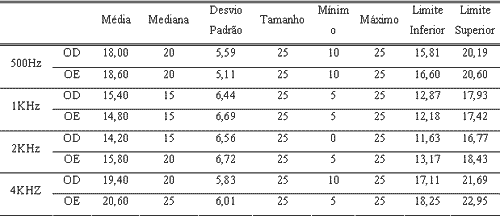
Table 1 shows descriptive analysis of audibility thresholds of patients with normal auditory sensitivity that comprised the studied group.
Duration Pattern Test (TPD) and Frequency Pattern Test (TPF) were presented in this order, monoaurally, in soundproof booth, with audiometer model AC33, brand Interacoustics, calibrated according to norm ANSI 69.
We used compact CD, reproduction of Speech and Pure Tone Material for Auditory Perceptual Assessment, disk 1.0 1994, recording made in professional studio with digital recording equipment. The test was presented at 50dBSL, that is, 50dB above the mean of frequencies 500, 1000 and 2000Hz, and patients were expected to respond verbally through naming 11.
Frequency patterns comprised three tones of 150ms and two 220ms-intervals of sounds. The tones, in each frequency pattern, were combinations of two sinusoid curves, 880Hz and 1,122Hz, designated as low frequency (G) and high frequency (A). There were six possible combinations of three tones in sequence, with two repetitive tones and one different one (GGA, AAG, GAG, AGA, GAA, AGG). Tones were generated digitally and there were 60 sequences, with approximately six seconds of interval in between them, and 30 sequences were presented on the right ear and 30 on the left ear.
Duration patterns comprised three tones of 1000Hz and two intervals between tones of 300ms. The tones in each duration pattern had either 250ms or 500ms and were designated as short (C) or long (L), respectively. The tones were digitally generated and their combinations resulted in six patterns (LLC, CCL, LCL, CLC, LCC, CLL), which were repeated equally and randomly ten times each one, for which the expected answer was naming. The number of presented sequences was 60, being 30 in each ear.
Standardization to the Brazilian population in tests of Duration Pattern and Frequency Pattern were made by Corazza (1998), in which they reached the indexes of correct answers, considering the variation range between percentile 3 and 97, as follows: TPF test values between 76 and 100% of correct answers, and TPD test of 83 to 100% of correct answers. As to mean number of correct answers, for TPD test they found 95.87% of correct answers and for TPF test the value of correct answers was 91.27%.
To analyze the results we used the following statistical techniques: Anova, Correlation (correlation matrix implies that 0% to 20% - bad correlations, 21% to 40% - poor correlations, 41% to 60% - fair correlations, 61% to 80% - good correlations, and 81% to 100% - excellent correlations), Confidence interval and equality of two proportions, in addition to Pearson's correlation (determines to what extent variables are interconnected, that is, how much they are related). The results were presented in percentage.
RESULTSInitially, we compared performance obtained in Frequency and Duration Pattern Tests with variable side of the ear (right or left). Results found are described in Table 2.
We observed that there were no statistically significant differences in Frequency and Duration Pattern Tests according to variable side of ear.
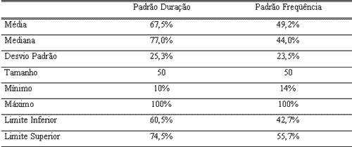
Thus, we gathered data obtained from both ears and conducted the descriptive analysis of correct answers obtained in frequency and duration pattern tests to a new composition (N=50 ears). Results are presented in Table 3.
Next, we correlated the results obtained with Duration and Frequency Pattern Tests and age of patients that comprised the studied group (Table 4).
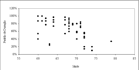
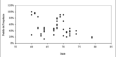
We correlated age and results found in Frequency and Duration Pattern tests and observed regular correlation between them, being that they were inversely proportional, that is, as age increased, percentage of correct answers decreased. The highest correlation was seen in the Duration Pattern Test (50.9%).
Results of these correlations are shown in graphic form in Figures 1 and 2.
DISCUSSIONElderly may present auditory processing disorders even without structural lesions that can directly explain them.
Authors have tried to characterize and determine the neural bases of presbyacusis and demonstrated the effect of aging plus peripheral hearing loss, which implied a conjunction of central dysfunction, even without structural damage and peripheral hearing loss in presbyacusis. These two added factors bring serious problems to the daily lives of elderly subjects, hindering speech recognition in quiet and especially in noise 13.
Many studies have been made in order to assess deficits caused by aging in auditory processing. Research studies pointed to the fact that aging can lead to progressive brain asymmetry, causing reduction of auditory information, which can be explained by inefficiency of inter-hemispheric transfer of information 5, 14-16.
Other studies tried to associate results of different tests trying to find a possible correlation between them. In the study that analyzed P300, pure tone audiometry and auditory processing in the elderly it was observed that P300 was intact in patients with auditory processing disorders, because latency and amplitude of potential in the elderly did not suffer influence of gender, nor of assessed central derivations. Abnormalities observed in P300 can only be related to systemic diseases and abnormalities in pure tone audiometry. It suggests that alterations in auditory processing are not necessarily accompanied by structural lesions of central nervous system 6, 4. These results have also shown that auditory processing disorders may not always be correlated with presence of peripheral hearing loss.
Many different authors emphasized the importance of sound temporal sequence perception for the identification and discrimination of speech sounds, in addition to emphasizing the importance of inter-hemisphere integration for this skill 17-19. Much has been said about the importance of ordering and temporal sequencing in auditory system, being that they are basic functions for speech discrimination and interpretation. Authors also emphasized the role of hemispheric integration and the participation of other areas of the central nervous system to conduct this task 7. Thus, in order to study these auditory skills, Frequency Pattern test and Duration Pattern test have been used to assess temporal auditory processing 20.
Some authors stated that the difficulties to understand speech in the elderly cannot be associated only with peripheral hearing loss. They specifically studied auditory temporal aging and observed that there are differences in temporal resolution between young and elderly, which are not explained only by the presence or absence of auditory loss 21.
Some studies in young Brazilian adults with normal hearing have also been conducted by the application of Duration Pattern tests (TPD) and Frequency Pattern tests (TPF), to standardize and allow clinical use 11.
The present study applied Duration Pattern and Frequency Pattern tests (TPD and TPF) in elderly with normal hearing sensitivity and we observed there was no influence of side of ear (right or left) (Table 2). Thus, data from both ears were grouped and mean index of correct responses found in the tests of Duration and Frequency, respectively, were 67.5% and 49.2% (Table 3).
As to variable side of ear, results found in this study agree with the findings by other authors 11, 20, 22, 23, in which we observed that there was no interference of side of ear.
As to mean of correct responses found for Duration and Frequency Pattern Tests in the elderly, we observed significant reduction in the percentage of correct responses when compared with young subjects with normal hearing sensitivity 9, 11, 20. These findings agree with those found in another study 24 that has also assessed auditory processing in the elderly, detecting values below those expected for young adults.
The results can be explained by the influence of aging in auditory processing, as previously addressed. In a study designed to assess development and maturation of central auditory system, using Duration and Frequency Pattern tests with subjects aged 7 to 16 years, it was observed progressive improvement in scores of tests, as age increased 12. Upon studying interaural differences in many hearing tasks in elderly and young people, the authors came up with the hypothesis that hemispheric asymmetry would be attenuated by development, being minimum in young adults and reappearing as a result of the decline of the auditory function caused by aging; it could be related with progressive loss of efficiency in inter-hemispheric transfer of auditory function, which is closely related with ordering and temporal sequencing skills and can be observed in the results of Duration Pattern and Frequency Pattern tests 14. However, it is important to highlight that these tests are not influenced by side of ear to which the stimulus is presented, which can be related with the hypothesis that the right and left hemispheres work together to conduct this task 7, 8.
Thus, the results obtained with this study suggested that elderly subjects with normal hearing present normal standards below that of young subjects with normal hearing. However, new studies should be conducted in order to define new standardization for Duration Pattern and Frequency Pattern tests in the elderly.
CONCLUSION- There is no difference in performance in Frequency Pattern and Duration Pattern Tests according to variable side of ear (right and left).
- Elderly subjects with normal auditory sensitivity present average percentage of correct answers of 49.2% in the Frequency Pattern Test and 67.5% in the Duration Pattern Test, which are results inferior to those obtained in normal hearing young adults 11.
REFERENCES1. Pereira LD. Avaliação do Processamento Auditivo. In: Lopes Filho O. Tratado de Fonoaudiologia. São Paulo: Roca; 1997. p.109-26.
2. ASHA. Guidelines for Hearing Aid Fittings for Adults. American Journal of Audiology 1996: 41.
3. Carvallo RMM. Processamento Auditivo: Avaliação Audiológica Básica. In: Pereira LD, Schochat E. Processamento Auditivo Central: Manual de avaliação. São Paulo: Lovise; 1997. p.27-35.
4. Baran JA, Musiek FE. Behavioral Assessment of the Central Auditory Nervous System. In: Musiek FE, Rintlmann WF. Contemporary Perspectives in Hearing Assessment. Sydney: Allyn and Bacon; 1999.
5. Jerger J, Jeger S, Oliver T. Perozollo, F. Speech understanding in the elderly. Ear and Hearing 1989; 10: 79-89.
6. Nunes FB. Da Avaliação do P300 e do Processamento Auditivo em Pacientes Idosos com e Sem Queixas Auditivas [tese]. São Paulo: Universidade Federal de São Paulo; 2002.
7. Pinheiro ML, Musiek FE. Sequence and temporal ordering in the auditory system. In: Assessment of central auditory dysfunction - foundation and clinical correlates. Baltimore: Willians & Wilkins; 1985. p.219-38.
8. Musiek FE, Pinheiro ML. Frequency Patterns in Cochlear, Brainstem and Cerebral Lesions. Audiology 1987; 26: 79-88.
9. Musiek FE, Baran JA, Pinheiro ML. Duration Pattern Recognition in Normal Subjects and Patients with Cerebral and Cochlear Lesions. Audiology 1990; 29: 304-313.
10. Castro LC. Avaliação do processamento auditivo central em indivíduos com lesão cerebral: teste de padrão de duração [tese]. São Paulo: Universidade Federal de São Paulo; 2001.
11. Corazza MCA. Avaliação do Processamento Auditivo Central em adultos: testes de padrões tonais auditivos de freqüência e teste de padrões tonais auditivos de duração [tese]. São Paulo: Universidade Federal de São Paulo; 1998.
12. Schochat E, Rabelo CM, Sanfins MD. Processamento Auditivo Central: Testes Tonais de Padrão de Freqüência e de Duração em Indivíduos Normais de 7 a 16 anos de idade. Pró-Fono Revista de Atualização Científica 2000; 12(2):1-7.
13. Frisina DR, Frisina RD. Speech recognition in noise and presbycusis: relations to possible neural mechanisms. Hearing Research 1997; 106: 95-104.
14. Jerger J. Functional asymmetries in the auditory system. An Otol Rhinol Laryngol 1997; 6: 23-30.
15. Greenwald RR, Jerger J. Aging Affects hemispheric Asymmetry on a Competing Speech Task. J Am Acad Audiol 2001; 12: 167-173.
16. Jerger J, Estes R. Asymmetry in Event-Related Potentials to Simulated Auditory Motion in Children, Young Adults, and Senior. J Am Acad Audiol 2002; 13: 1-13.
17. Hirsh IJ. Auditory perception of temporal order. J acoust Soc Am 1959; 31: 759-67.
18. Swisher l, Hirsh IJ. Brain damage and the ordering of two temporally successive stimuli. Neuropsychologia 1972; 10: 137-52.
19. Pinheiro ML. Auditory pattern perception in patients with left and right hemisphere lesions. Ohio J. Speech. Hear. 1976;12: 9-20.
20. Musiek FE. Frequency (Pitch) and duration pattern tests. J Am Acad Audiol 1994; 5:265-8.
21. Neves VT, Feitosa MA. Controvérsias ou complexidade na relação entre processamento temporal Auditivo e envelhecimento. Rev Bras Otorrinolaringol 2003; 69 (2): 242-9.
22. Tillman G, Jerger J. Temporal Compounds Reveal Interaural Biases. J Am Acad Audiol 2002; 13: 285-294.
23. Zanoni LG. Processamento Auditivo Central em Idosos: Teste de Padrão de Freqüência e Duração. São Paulo: Universidade Federal de São Paulo; 1999.
24. Sanches ML. Avaliação do Processamento Auditivo em Idosos que relatam ouvir bem [tese]. São Paulo: Universidade Federal de São Paulo; 2002.
Table 1. Characterization of the sample according to audibility thresholds obtained in pure tone audiometry.

Table 2. Comparative study of correct responses obtained in the Duration Pattern Test (TPD) and Frequency Pattern Test (TPF) according to variable side of ear.

Table 3. Descriptive analysis of percentage of correct responses obtained in Duration Pattern Test and Frequency Pattern Test.

Table 4. Study of the correlation between age and percentage of correct responses in the Duration Pattern Test (TPD) and Frequency Pattern Test (TPF) with Pearson's correlation test.

 Figure 1.
 Correlation between percentage of correct responses and variable age in the Duration Pattern Test.
 Figure 2.
 Correlation between percentage of correct responses and variable age in the Frequency Pattern Test.


