INTRODUCTIONEver since myringoplasty was performed by Zöllner1 and Wullstein2 in 1952, several types of material have been used as graft to close tympanic membrane (TM) perforations, such as vein3, fascia4, fat5 and perichondrium6. Among the group, temporal muscle fascia and perichondrium are used with great success. On the other hand, cartilage use in middle ear surgery has been reported; it has accurate indications and some advantages over the other materials, especially in cases of atelectasic otitis, cholesteatomatous otitis and revision myringoplasty, despite the controversies of outcome regarding hearing capabilities in these cases due to cartilage graft's features6.
The use of cartilage for closure of TM perforations through transcanal access and inlay technique was first described by Eavey in 1998. The technique shows excellent results to close small or medium non-marginal perforations, with advantages such as easy technique, high speed and postoperative comfort to the patient7.
Despite its great applicability and success reported in the first results, more cases and follow-up reports of patients should be published for long-term evaluation of the graft, occurrence of failures, and possible causes, and standardization for technique indication regarding age of the patient and perforation size.
In Brazil, Lubianca-Neto et al. reported the first results in 1998, with 100% of graft take-rate in their cases and hearing improvement in all of them8. Subsequently, with 20 case reports, they showed 90% success rate of graft take-rate and evaluated the hearing results from surgeries9. They also conducted randomized studies for the inlay technique with cartilage and the conventional technique with temporal muscle fascia, making tympanum-meatus flap. The results showed no statistically significant differences, despite the great benefits of using inlay cartilage, namely: less postoperative care, lower cost for the health system, low morbidity and greater comfort to the patient10.
This study shows the outcome of inlay myringoplasty using tragus cartilage according to the principles of Eavey's technique in patients who underwent surgery over the last three years at the Department of Otorhinolaryngology, Hospital das Clínicas, Medical School, Ribeirão Preto, University of Sao Paulo.
OBJECTIVEAnalysis of the outcome of inlay myringotomy with tragus cartilage according to Eavey's technique, focusing on the graft take-rate and hearing improvement, through pre and postoperative audiometry, in patients who underwent surgery at the Department of Otorhinolaryngology, Hospital das Clínicas, Medical School, Ribeirão Preto, University of Sao Paulo.
MATERIAL AND METHODSPatients
In the present study, we evaluated 32 surgeries performed between December 2001 and February 2003 at the Department of Otorhinolaryngology of Hospital das Clínicas, Medical School, Ribeirão Preto, University of Sao Paulo. All patients who were not submitted to postoperative audiometry were not included in this study. Five patients had surgery in both ears and each ear was considered an individual case. Patients' age varied from 11 to 53 years. One patient had preoperative profound sensorineural hearing loss. Nine cases had undergone previous myringoplasty using the conventional underlay technique, with temporal muscle fascia.
The size of TM perforation varied between 2mm and 5mm in diameter, where perforations of up to 3mm were considered small and from 3 to 5mm, moderate. All patients were diagnosed with uncomplicated chronic otitis media and the middle ear was healthy. Pre and postoperative audiometry was performed in all patients considering gap average (difference between thresholds through air and bone conduction) in 500Hz, 1000Hz and 2000Hz. Gap average of less than 30 dB of hearing sensitivity was considered an inclusion factor, an important indication for myringoplasty.
The following parameters were used for the inlay technique: perforation size (up to 5mm), visualization of the whole perforation, uncomplicated chronic otitis media, with healthy middle ear, and no external ear irregularities and sinuosity, which could affect the endoaural approach.
Procedure
All surgeries were performed under general anesthesia and orotracheal intubation.
After positioning the patient, the tympanic membrane was microscopically observed, the outer ear was carefully cleaned and a delicate scarification was performed on the perforation borders. Subsequently, the perforation size was measured with a 90 degrees hook, 2mm wide. All surgeries were performed through transcanal approach.
The tragus cartilage graft was removed after a 1.0 cm incision on tragal dome and cartilage dissection from adjacent tissue; the perichondrium was maintained in both sides. Cartilage removal did not affect the tragus support. Skin was apposed with simple interrupted sutures, nylon 5-0.
Based on the TM perforation size, the cartilage graft was cut on a circular shape, with borders about 2mm larger than the perforation. Perichondrium was maintained in only one of the sides of the graft. A medial incision was made around the graft, so as that one could see two cartilage sheets bound by the middle, and one of them was recovered by perichondrium.
The middle ear cavity was filled with gel foam up to 2mm from the perforation. Then, the graft was placed, under microscopic visualization, over the perforation, using a technique similar to insertion of a ventilation tube. The final result showed the cartilage sheet without perichondrium placed laterally to the perforation, whereas the cartilage sheet recovered by perichondrium was placed medially to the perforation. Perichondrium was used to cover the graft borders. The whole perforation was checked. No skin graft from arm skin was placed over the cartilage, as initially described by Eavey4.
Postoperative care was provided using an earplug during the first day, avoiding wetting the ear during the first month and prophylactic antibiotic treatment with amoxicillin for 7 days. When mucous or purulent discharge was noted during the first week after surgery, antibiotic drops were prescribed for 10 days.
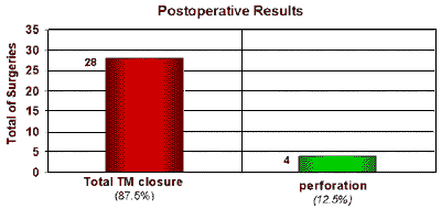
RESULTSOne month after surgery, total healing of TM perforations was accomplished in 28 of 32 surgeries performed, with 87.5% success rate during postoperative follow-up of 4.7 years (Chart 1). In cases of total take-rate, all grafts were intact and dry over the last visit (Figure 1). There was no significant postoperative complication. There was no report of problems on the skin incision on the tragus.
The surgeries were performed by resident physicians and medical professors and assistant professors from the Department of Otorhinolaryngology, Hospital das Clínicas, Medical School, Ribeirão Preto, University of Sao Paulo. Although the technique performed was standardized, the surgeons were not.
Out of the total cases of surgical success (28 patients), nine had moderate perforations (3-5mm) and 19 had small perforations (up to 3 mm).
In pre and postoperative audiometry in cases of total graft take-rate, we noticed the following results: nine patients had no preoperative gap and 1 had severe hearing loss; all of them had no audiometry changes after surgery. Eighteen patients had preoperative gap between 5 and 30 dB. In the postoperative audiometry, only 3 patients had no hearing improvement, the remaining (15 cases) showed average improvement of 14.4 dB (Chart 2).
In the cases of surgical failure (4 cases), only 1 had postoperative audiometric improvement (10 dB) (Chart 3).
DISCUSSIONThe use of cartilage in ear surgery has been widely studied and described, especially in advanced cases of middle ear diseases, such as revision cases, severe retraction of TM, atelectasia, cholesteatoma or perforation of auditory tube. Results of audiometry after using this graft have also been described. There were no significant differences in comparison with the use of other material (e.g., temporal muscle fascia). Therefore, cartilage graft is a good choice for middle ear disease management6.11.
Upon approaching uncomplicated chronic otitis media, when the middle ear is healthy, Eavey described his inlay technique using tragus cartilage shaped like butterfly wings to close TM perforations, with 100% success rate7. In Brazil, the results of this technique showed by Lubianca-Neto et al. achieved graft take-rate of 100% and 90%, respectively, on both studies conducted by them8,9.
The present study accomplished high success rate in closing TM perforations (87.5% of the cases). The principles were followed, with few changes to the technique, namely: in the group of patients, adults were included, the middle ear was filled with gelfoam before placing the graft, providing extra support and stabilization to the cartilage, the perichondrium was only maintained at the graft's medial sheet, reducing cartilage thickness and aiding its placement and maintaining its viability12; skin graft was not used over the cartilage, reducing morbidity and time of procedure, without affecting the results. The changes above-mentioned aimed at improving and adjusting the original technique according to our service expertise to maintain high success rate of the procedure.
Although the average postoperative follow-up period was short, we noted that the cartilage adhered to the tympanic membrane and was nourished through diffusion, a feature of this material. This is an avascular material, resistant to hostile environments that are poorly irrigated for long periods13.
The success to close TM perforations also reduced the postoperative gap in 83% of the cases of preoperative conductive hearing loss. In other cases of surgical success, the audiometric thresholds did not change. We noted that there was no influence affecting the audiometric results when we used cartilage as TM graft, and most of the cases showed improvement as provided by studies previously described7,8,9,10. We believe that the thinner the graft, the lesser the chance to affect (or not improve) the patient's hearing capabilities. To prove that, we removed the perichondrium from one side of the graft.
According to the original description, we also noted advantages using the inlay technique with tragus cartilage because the procedure is faster, there is no need to manufacture a meatal flap or any other incision on the outer ear, the outer ear does not need specific care or support and the graft is easily obtained. All of that provides better comfort to the patient with highly satisfactory results, similarly to the conventional technique (with temporal muscle fascia)7,10.
This technique is contraindicated in cases requiring middle-ear exploration, such as imbalance between conductive hearing loss and the perforation size, possible cholesteatoma or ear discharge, marginal perforations without visible borders, perforations that cannot be totally visualized through transcanal approach, and granulomatous process affecting tympanic membrane 7.
Although different surgeons, with different skills and expertise, performed the surgeries in this study, the procedure was technically simple and graft stability after correct placement was essential to the success rate of 87.5%, provided that the patient was correctly prepared for surgery. Therefore, we believe that the 4 unsuccessful cases of this study resulted from bad anchorage of the entire graft on the borders of the perforation and from infection at early postoperative period, non-responsive to topical antibiotics.
CONCLUSIONSThis study showed that inlay myringoplasty with tragus cartilage shaped like butterfly wings has high success rate in closing tympanic membrane perforations of up to 5mm in diameter in cases of uncomplicated chronic otitis media, when we can visualize all perforation borders, the middle ear is healthy and there is no need to explore the middle ear or carry out any procedure on the ossicle chain.
This procedure improves hearing capabilities in most of the patients who present conductive hearing loss at preoperative evaluation.
Since the technique is easily performed, fast, the access is through transcanal, without incision, and it is easy to obtain and shape the graft, Eavey's surgery and its present variants described are a good choice for myringoplasty in cases of uncomplicated chronic otitis media, with the features reported previously, and they provide more comfort to the patient and less morbidity.
REFERENCES1. Zöllner F. The principles of plastic surgery of the sound-conducting apparatus. J Laryngol Otol 1995; 69: 657-9.
2. Wullstein HL. Funktionelle Operationen im Mittelohr mit Hilfe des Freien Spaltlappentransplantates. Arch Otorhinolaryngol 1952; 161: 422-35.
3. Tabb HG. Closure of perforations of the tympanic membrane by vein grafts: a preliminary report of 20 cases. Laryngoscope 1960; 70: 271-4.
4. Herman H. Tympanic membrane plastic repair with temporalis fascia. Hals Nas Ohrennh 1960; 9: 136-9.
5. Ayache S, Braccini F, Facon F, Thomassin JM. Adipose graft: an original option in myringoplasty. Otol Neurotol 2003 Mar; 24(2): 158-64.
6. Dornhoffer JL. Hearing Results With Cartilage Tympanoplasty. Laryngoscope 1997; 107(8): 1094-9.
7. Eavey RD. Inlay Tympanoplasty: cartilage butterfly technique. Laryngoscope 1998 May; 108(5): 657-61.
8. Lubianca Neto JF. Miringoplastia com Cartilagem Inlay: Primeiros Resultados Brasileiros. Revista Brasileira de Otorrinolaringologia 2000; 66 (4): 310-4.
9. Lubianca-Neto JF. Inlay butterfly cartilage tympanoplasty (Eavey technique) modified for adults. Otolaryngol Head and Neck Surg 2000 Oct; 123(4): 492-4.
10. Mauri M. Evaluation of inlay butterfly cartilage tympanoplasty: a randomized clinical trial. Laryngoscope 2001; 111(8): 1479-85.
11. Gerber MJ, Mason JC, Lambert PR. Hearing results after primary cartilage tympanoplasty. Laryngoscope 2000 Dec; 110(12): 1994-9.
12. Smyth GDL, Kerr AG. Cartilage homografts. Experimental and clinical aspects. Acta Oto-rhino-laryngologica belg, 1970; 24: 53-9.
13. Levinson RM. Cartilage-perichondral composite graft tympanoplasty in the treatment of posterior marginal and attic retraction pockets. Laryngoscope 1987; 97: 1069-74.
 FIGURE 1-
 Appearance of the tympanic membrane after surgery.
 GRAPH 1-
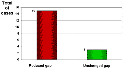 GRAPH 2-
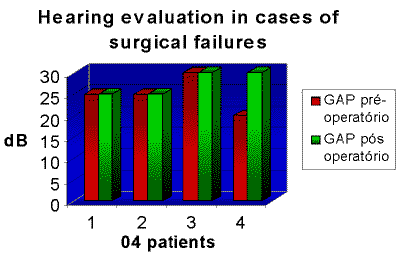 GRAPH 3-