IntroductionMeniere’s disease, an inner ear affection that is clinically manifested by fluctuating hearing loss, vertigo and tinnitus, whose pathophysiological substrate is endolymphatic hydrops, has been subject to a number of studies and controversial issues since its first description by Prosper Menière in 18611.
In 1995, the Committee on Hearing and Equilibrium Guidelines of the American Academy of Otorhinolaryngology and Head and Neck Surgery (AAO-HNS) defined clinical criteria for the diagnosis of Meniere’s disease with four levels of certainty (absolute, defined, likely, possible) and audiological criteria2. However, we found a number of studies with objective methods to study this disease, especially electrocochleography and otoacoustic emissions3-8.
Transtympanic electrocochleography has been considered the golden standard in objective diagnosis of endolymphatic hydrops, with a sensitivity of 83%4, using measures such as summation potential and action potential (PS/PA) using an alternate click to cancel cochlear feedback, with values ranging from 0.33 to 0.501, 3, 4, 5, 6, 9. Levine (1992)10, Johansson (1997)5 and Sass (1998)4,6 studied the latency of wave I using condensation and rarefaction clicks and showed statistically significant differences in patients with Meniere’s disease compared to normal subjects or those with other cochlear-vestibular affections.
The present study aimed at assessing wave I latency in transtympanic electrocochleography using condensation and rarefaction clicks in a group of patients with clinical diagnosis of Meniere’s disease and the difference between the values, considering the results in the relation between summation potential and action potential.
Material and MethodWe retrospectively assessed the clinical charts of patients who underwent transtympanic electrocochleography in the Otolaryngology Diagnostic Center in Florianópolis-SC, from December 2000 to April 2002. We selected 22 patients (22 ears) with likely clinical diagnosis of Meniere’s disease according to the criteria of AAO-HNS2. The data collected were age, gender, mean thresholds for pure tone audiometry in frequencies of 500, 1000, 2000 and 3000 Hz in the ear evaluated by electrocochleography, the PS/PA ratio in tracings of alternate clicks, wave I latency using rarefaction and condensation alternate clicks and the difference between latency with the use of one and the other click.
All electrocochleography exams were performed by the author of this study using a needle electrode as the active electrode, introduced through the tympanum, previously anesthetized with phenol at 89%, and disk electrodes on the forehead and ipsilateral mastoid region as reference and grounding, respectively. The stimuli used were alternate polarity clicks, condensation and rarefaction, of up to 11/s rate, duration of 100ms, summation of 1,000 tracings and intensity of 120dB SPL. We considered the value of the PS/PA ratio greater or equal to 0.339 as an indication of endolymphatic hydrops and used the parameter to divide patients in the two groups and to compare the results of data collected from them.
ResultsWe assessed 22 patients, 16 female and 6 male subjects, and in 8 cases (36.4%) the PS/PA ratio was greater or equal to 0.33, defined as group 1, and in 14 cases (63.6%) PS/PA ratio was below 0.33, defined as group 2. The ratio ranged from 0.11 to 0.78. The mean age of group 1 was 46.4 years and group 2, 44.4 years.
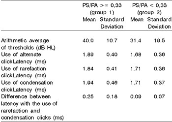
The latency measures with alternated rarefaction and condensation clicks, the difference in latencies using condensation or rarefaction clicks and arithmetic average of thresholds with standard deviations for both groups are shown in Table 1.
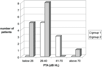
The distribution of patients and hearing loss is presented in Graph 1.
Table 1. Results of audiometric exams and wave I latency according to PS/PA.


Graph 1. Distribution of Patients in Relation to Severity of Hearing Loss. PTA: Arithmetic average of frequencies 500, 1000, 2000 and 3000 Hz.
The assessed patients presented similar characteristics to those presented in other studies. There was predominance of female patients and the mean age range was 44 to 46 years, similarly to the study by Levine (1998)3. Celestino (1991)11 and Green (1991)12 found mean age of 48 years. Thus, our sample was representative concerning age and gender.
The analysis of PS/PA ratio showed 36.4% of the patients with values greater or equal to 0.33, indicating endolymphatic hydrops. The number of patients with abnormal PS/PA ratio is variable in the literature. Ferraro (1985)13 found 36 patients with abnormalities in 110 clinical diagnoses of Meniere’s disease, whereas Goin had 62% in his study14. Conlon (2000)1, using the cutoff criterion of PS/PA ratio greater or equal to 0.50, found 30% of the patients with abnormal results. It has been argued that the positive result of electrocochleography in the diagnosis of Meniere’s disease and PS/PA ratio would be greater in early stages of the disease and when the patient is approaching a crisis, which would justify the differences in values found among authors since the patients were not homogenous in many studies3. Levine (1998) found a greater number of subjects with abnormal PS/PA ratio when there was history of Meniere’s disease of 7 to 12 months compared to those with longer history of the disease3.
It is known that endolymphatic hydrops leads to distension of basilar membrane, modifying the sound transmission mechanics in the inner ear, which is one of the explanations for the abnormal amplitude of summation and action potentials at electrocochleography. Similarly, since there is distension of the basilar membrane, the transmission of the travelling sound wave with the use of condensation and rarefaction clicks would be different, resulting in a difference of wave I latency as a response to them4. Sass4,6 and Johansson5 used condensation and rarefaction clicks and compared the results between patients with Meniere’s disease and other cochlear-vestibular diseases or normal subjects and found an increase in wave 1 latency when using the condensation clicks and differences in the values of wave I latency when using the rarefaction and condensation clicks in the group of Meniere’s disease patients compared to the others. Levine, in his study, defined that if the difference between such latencies was greater or equal to 0.30ms it was an indication of endolymphatic hydrops 10. In our study, we used a condensation and alternate click, but compared the results between patients with clinical diagnosis of Meniere’s disease, but separating them according to the results of the PS/PA ratio, considering that the greater than 0.33 the value, the more endolymphatic hydrops would be present, together with basilar membrane abnormal position and the wave transmission described above. Thus, we could evaluate whether wave I latency values with different stimuli could reflect the mechanic abnormalities of the endolymphatic duct and whether they could be used as a parameter for the diagnosis of endolymphatic hydrops. We found that the mean wave I latency using the condensation click was greater than the latency with the rarefaction click between the studied groups, but it was not statistically significant, differently from other authors reported here, but our sample was very small. Similarly, the difference in latencies was present in group 1 (PS/PA>=0.33), but not significantly, being below the value of 0.30ms established by Levine10, closer to the value of 0.23ms found by Saas5, but we strongly believe that the size of the sample influenced the result. This value may seem to be paradoxical since the latencies did not present statistically significant differences, but they represent averages and in some patients the values for latency with rarefaction click were greater than for condensation clicks, which reflected the differences in absolute value calculations.
Sass, considering the value of the referred difference in latency, detected increase in sensitivity of electrocochleography for the diagnosis of Meniere’s disease from 83 to 87%, maintaining the specificity of almost 100%, but the data isolated resulted in 37% of sensitivity only. However, in cases in which it is difficult to have the PS/PA ratio, it can help with the diagnosis, since it has high specificity4.
During the conduction of the exams, the use of rarefaction and condensation clicks, in addition to the alternate clicks, did not result in significant increase of duration of the test, and it was easy to perform. As discussed, it may add further data that can increase sensitivity to electrocochleography in Meniere’s disease diagnosis.
ConclusionsIn patients in group 1, the values of wave I latency with condensation clicks and the difference in latency with condensation and rarefaction clicks was greater than in group 2, but they were not statistically significant differences. The assessment of these parameters should be further investigated so that we can clearly define their importance in clinical practice.
References 1. Conlon BJ, Gibson WPR. Electrocochleography in the Diagnosis of Meniere’s Disease. Acta Otolaryngol 2000;120(4):480-483.
2. Committee on Hearing and Equilibrium Guidelines for the Diagnosis and Evaluation of Therapy in Menière’s Disease. Otolaryngol Head Neck Surg 1995;113(3):181-185.
3. Levine S, Margolis RH, Daly KA. Use of Electrocochleography in the Diagnosis of Meniere’s Disease. Laryngoscope 1998;108(7):993-1000.
4. Sass K, Densert B, Magnusson M, Whitaker S. Electrocochleographic Signal Analysis: Condensation and Rarefaction Click Stimulation Contributes to Diagnosis in Menière’s Disorder. Audiology 1998;37(4):198-206.
5. Johansson RK, Haapaniemi JJ, Laurikainen EA. Transtympanic Electrocochleography in Evaluation of Cochleovestibular Disorders. Acta Otolaryngol (Stockh) 1997; suppl 529:63-65.
6. Sass K, Densert B, Arlinger S. Recording Techniques for Transtympanic Electrocochleography in Clinical Practice. Acta Otolaryngol (Stockh) 1998;118(1):17-25.
7. Sakashita T, Kubo T, Kusuki M, Kyunai K, Ueno K, Hikawa C et al. Patterns of Change in Growth Function of Distortion Product Otoacoustic Emissions in Meniere’s Disease. Acta Otolaryngol (Stockh) 1998;Suppl 538:70-77.
8. Kusuki M, Sakashita T, Kubo T, Kyunai K, Ueno K, Hikawa C, et al. Changes in Distortion Product Otoacoustic Emissions from ears with Meniere’s Disease. Acta Otolaryngol (Stockh) 1998;Suppl 538:78-89.
9. Gibson WPR, Prasher DK, Kilkenny GPG. Diagnostic Significance of Transtympanic Electrocochleography in Menière’s Disease. Ann Otol Rhinol Laryngol 1983;92:155-159.
10. Levine SC, Margolis RH, Fournier EM, Winzenburg SM. Tympanic Electrocochleography for Evaluation of Endolymphatic Hydrops. Laryngoscope 1992;102:614-22.
11. Celestino D, Ralli G. Incidence of Meniere’s Disease in Italy. Am J Otol 1991;12(2):135-138.
12. Green JJ, Blum DJ, Harner SG. Longitudinal Follow Up Patients with Meniere’s Disease. Otolaryngol Head Neck Surg 1991;104(6):783-788.
13. Ferraro JA, Arenberg IK, Hassanein RS. Electrocochleography and Symptoms of Inner Ear Dysfunction. Arch Otolaryngol 1985;111(2):71-74.
14. Goin DW, Staller SJ, Asher DL, Mischke RE. Summating Potential in Meniere’s Disease. Laryngoscope 1982;92(12):1383-1389.
1 Otorhinolaryngologist, Hospital Governador Celso Ramos, Former Resident of the Discipline of Otorhinolaryngology, Medical School, University of São Paulo - USP.
2 Ph.D., Professor of the Discipline of Otorhinolaryngology, Medical School, USP.
3 Master studies under course, Discipline of Pneumology, Federal University of Rio Grande do Sul, Otorhinolaryngologist, Hospital Governador Celso Ramos.
4 Master studies under course, Discipline of Otorhinolaryngology, Medical School, Ribeirão Preto, USP, Head of the Service of Otorhinolaryngology, Hospital Governador Celso Ramos, Florianópolis-SC.
Study conducted at the Otolaryngology Diagnostic Center, Florianópolis-SC.
Address correspondence to: Cláudio M. Y. Ikino – Av. Jornalista Rubens de Arruda Ramos, 2560
Centro Florianópolis SC – 88015-700
Tel/fax (55 48) 224.1111 – E-mail: claudioikino@hotmail.com
Article submitted on August 16, 2002. Article accepted on September 19, 2002


