INTRODUCTIONAccidents with foreign bodies in children, including the esophagus, are observed daily in ENT emergency rooms. Among them, coins are the most common foreign bodies, amounting to about 70% of the cases, most of them in the cervical esophagus1, 2, 3, 4.
The clinical picture is very variable. About 20% are symptomatic1, 4, and they may manifest gastrointestinal symptoms (dysphasia and vomiting) and respiratory symptoms (cough and dyspnea). The latter can be caused by compression of the foreign body or secondary esophageal dilation over the trachea, as well as edema of paraesophageal soft tissues in case of prolonged retention of the foreign body1. In most cases, parents observe the intake of the foreign body or are advised by the child about it. In other occasions, the foreign body is a radiological finding in chest or abdominal x-rays.
The diagnosis is confirmed by radiological exam, requiring mentalis-xipho-pubic incidence, which enables visualization of the whole digestive tract. In most cases, the removal is made by rigid esophagoscopy under general anesthesia.
Complications are rare and noticed in cases of prolonged foreign body retention1, 5, including esophageal perforation, mediastinitis, tracheoesophageal fistula, vascular fistula, extra-luminal migration of foreign body and formation of false diverticulum 1, 5, 11, 12. There may be also cases of iatrogenia, such as perforations during esophagoscopy. If early diagnosed, before they evolve to mediastinitis, they have good prognosis after the surgical procedure. In cases of mediastinitis, prognosis is more reserved. Congenital abnormalities of the esophagus can also predispose to complications10.
We intended to analyze an alternative to the removal by esophagoscopy under general anesthesia: the removal with Foley catheter under local anesthesia, in the hospital setting. We assessed efficacy, safety and cost of the method compared to esophagoscopy under general anesthesia.
MATERIAL AND METHODWe included 25 children with esophageal foreign body (coins) seen by the service of ENT, Hospital Municipal Souza Aguiar, a reference center for ENT foreign body in the state of Rio de Janeiro, between September 1996 and April 1998.
They were submitted to placement of urinary catheters (Foley) to try to remove the foreign body and we did not use any type of anesthesia to perform the procedures; in some cases, we lubricated the catheter with xilocaine gel, but it seemed to hinder the placement, since the catheter became slippery.
The following parameters were observed before the procedure:
• only coins and telephone tokens can be removed by this method, because they do not have cutting surfaces;
• the foreign body should be single;
• the foreign body should be located maximum at the middle third of the thoracic esophagus;
• there should be a maximum of 36 hours between ingestion and the procedure;
• radiological findings should not be removed with this method, because they are normally associated with inflammatory phenomena, since we do not know how long the foreign body has been there;
• absence of disease or previous esophageal surgery;
• the procedures can only be performed in the hospital, with the presence of the pediatrician and the anesthesiologist.
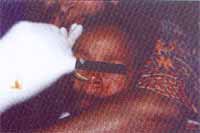
The procedure was explained in details to the child and the accompanying person. The catheters were introduced through the nose, the child was restrained by the escort (seating on his/her lap) supported by the licensed practical nurse (LPN).
After the introduction of the catheter, the balloon was inflated with air up to two thirds of its total capacity, and then it was pulled upwards to the oropharynx. The LPN flexed slightly the head of the patient and the physician introduced a gloved finger in the child’s oral cavity to help expel the foreign body, which eventually led to marks of bite in the gloved finger. After removal of the foreign body, the child remained in observation for 30 minutes and was then discharged.
When the foreign body was not eliminated, we performed up to three attempts. If there was no elimination yet, new x-rays were ordered to check the position of the foreign body. If the foreign body had regressed to the abdominal cavity, we sent the patient to the pediatrician. If it remained in the same site, we decided for esophagoscopy under general anesthesia.
We collected data such as age, gender, value of the coin, number of catheters used, interval between ingestion of coin and its removal, number of attempts (to pass the catheter), duration of procedure, complications and location of foreign body.
RESULTSOut of 25 cases, 15 (60%) were male and 10 (40%) were female subjects. Ages ranged from 1 to 13 years (mean of 5.1 years). The most frequent age range was 2 to 5 years (48%), followed by 6 to 10 years (32%).
As to location, 16 (64%) of the foreign bodies were in the inferior third of the cervical esophagus and 9 (36%) in the middle third of the thoracic esophagus. The most frequently found coins were, in order of frequency, R$0.01 (24%), R$0.05 (20%), R$0.10 (20%), R$0.25 (8%), R$0.50 (8%), old coins (8%) and R$1.00 (4%), curiously in decreasing order (the least valuable are also the smallest ones). In two cases (8%) we did not know the value because the coins progressed to the abdomen. The most frequently used catheters were No.10 and 12 (36% each), followed by No.14 (16%) and 16 (4%). In two cases, two catheters of different size were used. In 12 cases (48%), only one attempt was enough, seven cases (28%) required two attempts, and 6 cases (24%) three. As to interval between ingestion and removal, 9 cases (36%) were solved within the first 6 hours, 10 cases (40%) between 6 and 12 hours, 3 cases (12%) between 12 and 24 hours and 3 cases (12%) between 24 and 36 hours. The duration of the procedure ranged from 30 seconds to 10 minutes, in approximate values.
The foreign body was orally expelled in 20 cases (80%). In 4 (16%), it progressed to the abdominal cavity and in only one case (4%) the patient underwent esophagoscopy under general anesthesia.
In only one case (4%) there was a complication: mild bleeding through the oral cavity, and the patient was observed for 24 hours and remained uneventful.

Figure 1. Patient being teld for removal
The ingestion of coins by children and psychiatric patients is quite common. In a large number of cases, coins follow towards the stomach and are normally eliminated in the feces within some days. In other cases, they remain impacted in the esophagus, normally the cervical portion, preventing normal feeding and causing inflammatory processes that can lead to severe complications.
Our data were in accordance with the literature concerning age range (2 to 5 years), gender (60% of male patients), interval between ingestion and removal of foreign body (76% within the first 24 hours) and location of the foreign body (64% in the inferior third of the cervical esophagus).
In most emergency rooms, patients are submitted to rigid esophagoscopy under general anesthesia to remove the foreign body, which requires hospitalization and produces higher morbidity, resulting both from the endoscopic performance and the anesthetic act.
Foley catheters, normally used as urinary tubes, have been used for the removal of foreign bodies since the 60’s1. Initially, we force the progression to the stomach4, 6 and then extract it orally7. Many studies, especially in the North American literature, described the procedure guided by fluoroscopy 8, 9. In our opinion, this detail is dispensable, as agreed by some literature reports13,14,15,16,17,18,19.
In our center, we have been performing this procedure for a long time, observing high success rates (90% of the cases) and low rates of complications (4% of the cases) and none were severe. We emphasize the need for hospital infrastructure, with pediatrician and anesthesiologist on call, owing to the risk of bronchial-aspiration of the foreign body, even though we had never seen it happen, nor seen literature reports. The parameters previously listed should also be strictly respected, otherwise the rate of complications would be higher.
The mean length of hospital stay was very short (about 1 to 2 hours for admission, physical examination, x-ray, procedure and clinical observation), as opposed to the 12/24 hours required for esophagoscopy under general anesthesia, which reduced significantly the morbidity.
The total cost of material (Foley catheter, syringe and gloves) was at least 50 times cheaper than the expenditure with esophagoscopy under general anesthesia.
CONCLUSIONWe concluded that the method is safe, effective, quick and cheap to remove coins from the esophagus. Thus, by observing the protocol of restrictions, we strongly believe that the procedure should be at least tried, preventing the performance of esophagoscopy under general anesthesia in many cases and reducing morbidity significantly.
ACKNOWLEDGMENTWe would like to thank the whole team of otorhinolaryngologists and licensed practical nurses of Hospital Municipal Souza Aguiar.
REFERENCES 1. Macpherson IR et al. Esophageal foreign bodies in children. American Journal of Roentnology April 1996;166(4):919-24.
2. Nandi P, Ong GB. Foreign body in the esophagus: review of 2394 cases. Br Surg 1978;65:5-9.
3. Towbin R et al. Esophageal edema as a predictor of unsuccessful balloon extraction of esophageal foreign bodies. Pediatric Radiology 1989;19:359-360.
4. Emslander HC et al. Efficacy of esophageal bougienage by emergency physicians in pediatric coin ingestion. Annals of emergency medicine 1996 June;27(6):726-9.
5. Relly J et al. Pediatric aerodigestive foreign bodies injuries. Laryngoscope 1997 Jan;107(1);17-20.
6. Aiken DW. Coins in the esophagus: a departure from conventional therapy. Mil Med 1965;130:182-3.
7. Bigler FC. The use of a Foley catheter for removal of blunt esophageal foreign bodies from children. Thorac cardiovascular surgery 1966;51:759-760.
8. Shackelford GD et al. The use of a Foley catheter for removal of blunt esophageal foreign bodies from children. Radiology 1972;105:455-6.
9. Campbell JB, Davis WS. Catheter technique for extraction of blunt esophageal foreign bodies. Radiology 1973;108:438-440.
10. Hollinger PH et al. Congenital anomalies of the esophagus related to foreign bodies. Am Dis Child 1949;78:467-476.
11. Yee KF et al. Extramural foreign bodies (coins) in the food and air passages. Ann. Otolaryngology 1975;84:619-623.
12. Ramadan MF et al. An acquired esophageal pouch in childhood. J Laryn otology 1981;95:101-108.
13. Brown LP. Blind esophageal coin removal using a Foley catheter. Arch Surg 1968;96:931-2.
14. Spitz L. Management of ingested foreign bodies in childhood. BM1 1975;1:561-3.
15. Reully MS, Walter MA. Consumer products aspiration and ingestion: analysis on emergency reports. Ann Otolog Laryn 1992;101:109-111.
16. Harned RK et al. Esophageal foreign bodies: safety and efficacy of Foley catheters extraction of coins. AJR Am J Roentnology 1997;168(2):443-6.
17. Agarwala S et al. Coins can be safely removed from esophagus by Foley catheters without fluoroscopic control. Indian pediatric 1996;33(2):109-11.
18. Conners GP. A literature-based comparison of three methods of pediatric esophageal coin removal. Pediatric Emergency Care 1997;13(2):154-7.
19. Morrow SE et al. Balloon extraction of esophageal foreign bodies in children. J Pediatric Surg 1998;33(2):266-70.
1 Otorhinolaryngologist, Hospital Municipal Souza Aguiar, Rio de Janeiro.
2 Physician, Resident in Otorhinolaryngology, Hospital Municipal Souza Aguiar.
3 Trainee Physician, Hospital Municipal Souza Aguiar.
4 Trainee Physician, Hospital Municipal Souza Aguiar.
Study conducted at Hospital Municipal Souza Aguiar, Rio de Janeiro
Address correspondence to: Ricardo R. Figueiredo – Rua 60, nº 1680 ap. 202
Bairro Sessenta Volta Redonda RJ 27261-130
Fax (55 24) 3349-8664 –E-mail: otosul@uol.com.br.
Article submitted on May 23, 2002. Article accepted on July 11, 2002


